. 2012 Sep;30(9):2020-31.
doi: 10.1002/stem.1175.

Spatial domains of progenitor-like cells and functional complexity of a stem cell niche in the neonatal rat spinal cord

Affiliations

Spatial domains of progenitor-like cells and functional complexity of a stem cell niche in the neonatal rat spinal cord

Nicolás Marichal et al. Stem Cells. 2012 Sep.

Abstract

During spinal cord development, progenitors in the neural tube are arranged within spatial domains that generate specific cell types. The ependyma of the postnatal spinal cord seems to retain cells with properties of the primitive neural stem cells, some of which are able to react to injury with active proliferation. However, the functional complexity and organization of this stem cell niche in mammals remains poorly understood. Here, we combined immunohistochemistry for cell-specific markers with patch-clamp recordings to test the hypothesis that the ependyma of the neonatal rat spinal cord contains progenitor-like cells functionally segregated within specific domains. Cells on the lateral aspects of the ependyma combined morphological and molecular traits of ependymocytes and radial glia (RG) expressing S100β and vimentin, displayed passive membrane properties and were electrically coupled via Cx43. Cells contacting the ventral and dorsal poles expressed the neural stem cell markers nestin and/or vimentin, had the typical morphology of RG, and appeared uncoupled displaying various combinations of K(+) and Ca(2+) voltage-gated currents. Although progenitor-like cells were mitotically active around the entire ependyma, the proliferative capacity seemed higher on lateral domains. Our findings represent the first evidence that the ependyma of the rat harbors progenitor-like cells with heterogeneous electrophysiological phenotypes organized in spatial domains. The manipulation of specific functional properties in the heterogeneous population of progenitor-like cells contacting the ependyma may in future help to regulate their behavior and lineage potential, providing the cell types required for the endogenous repair of the injured spinal cord.


Conflict of interest statement

Disclosure of potential conflicts of interests:

The authors declare no conflicts of interests.

Figures
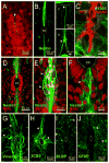
Figure 1
Figure 1. CC-contacting progenitor-like cells: molecular clues
A, Most cells surrounding the CC expressed the ependymal cell marker S100β. However, a narrow portion on the dorsal pole of the ependyma was devoid of S100β immunoreactivity (arrow). B, Nestin immunoreactivity was observed mostly in cells with long radial processes in the dorsal and ventral midline that stretched from the CC lumen (1) to the pia on the dorsal (2, arrowhead) and ventral (3, arrowhead) aspects of the cord. C, Nestin+ fibers on the dorsal CC did not co-express S100β. D, Location of nuclei (Syto64) belonging to nestin reactive fibers in the dorsal and ventral midline. E, Nestin+ cells on the dorsal raphe had a bipolar shape with their cell bodies located at various distances from the CC (arrowheads). F, The cell bodies of nestin+ cells were also found close to the CC lumen (arrowhead). G–H, RG markers vimentin and 3CB2 expressed in cells on the lateral aspects and poles of the ependyma, with some fibers projecting away from the CC (arrowheads). I, BLBP is not expressed in the ependyma but in cells outside the ependymal region (arrowheads). J, GFAP+ cells were not detected in the ependymal layer, but cells with the morphology of astrocytes appeared in the spinal cord outside this region (arrowheads). A–D, F and J, single confocal optical planes. E, G–H, confocal Z-stack projection. Unless otherwise stated, in this and subsequent figures the dorsal pole of the CC is upward.
Figure 2
Figure 2. Functional properties of cells lining the lateral CC: dye coupling and passive responses
A, DIC image of the CC in a live slice. B, Responses of a cell recorded on the lateral CC to a series of voltage steps (1) that displayed a linear I/V relationship (2). This cell lacked voltage-gated currents as shown in the leak subtracted traces (3). C, Distribution of input resistances and membrane potentials of cells recorded on the lateral aspects of the CC. D, The cell recorded in B appeared dye-coupled with neighboring cells. E, Carbenoxolone (100 μM) increased the input resistance of cells in the lateral aspects of the CC (n=5). F, Clustered cells covered the whole lateral aspect of the CC (1) or formed rather small cell conglomerates on the ventral (2) or dorsal (3) halves of the lateral CC. Some clustered cells had processes that entered the dorsal or ventral midline (2 and 3, arrowhead), projecting toward the pia (4, arrow). G, Cx43 expression in the CC. Cx43 punctae concentrated on the lateral aspects close to the CC lumen (arrow). D, Conventional epifluorescence in a living slice. F1-4, Confocal Z-stack projections. G, single confocal optical plane.
Figure 3
Figure 3. IKD and IA in midline RG
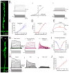
A, Alexa 488 filled RG contacting the ventral pole of the CC. B, Raw (1) and leak-subtracted (3) currents in a RG in response to a series of voltage steps. An outward current with an activation threshold of −40 mV (2) was generated in response to depolarizing voltage steps. C and D, Current amplitude and activation curves for IKD, respectively. E, The current was blocked by TEA (10 mM). F, RG recorded in the ventral midline. G, Currents evoked in the cell shown in F by depolarizing voltage steps after a prepulse to −90 mV (1). Currents with a slower onset were evoked with the same protocol as in 1 but after a pre-pulse to −30 mV (2). By subtracting 1 and 2, we obtained a current with a fast onset and voltage-dependent inactivation. H, IKD was blocked by TEA (10 mM, 1 and 2) whereas the remaining current was blocked by the IA antagonist 4-AP (2 mM, 2 and 3). I and J, IA activation/inactivation and amplitude curves, respectively. A and F: Confocal Z-stack projection. I–J: data pooled from P1–P5 rats.
Figure 4
Figure 4. ICa in midline RG
A, RG recorded and filled with Alexa 488. B, This cell displayed both outward and slow inward currents (1, arrow) in response to depolarizing voltage steps. The inward current remained in the presence of K+ channel antagonists (2, arrow, 4-AP 2 mM and TEA 10 mM) but was abolished in low Ca2+ Ringer solution (3), suggesting the involvement of voltage-gated Ca2+ channels. C, In current clamp mode, depolarizing current steps applied from a hyperpolarized membrane potential generated a slow spike (1, 2 arrow) that disappeared in low Ca2+ (2). A, Confocal Z-stack projection. .
Figure 5
Figure 5. Molecular and ultrastructural characteristics of cells contacting the dorsal pole of the ependyma
A, Nestin+ cells contained pericentrin+ granules in their apical processes (arrow). B, Electron micrograph of the apical processes of cells contacting the dorsal CC (inset). Each process had the insertion site of a single cilium (arrows). C, Longitudinal view of a cilium (arrow). Some cilia exhibited a 9 + 0 microtubule organization (inset, arrowhead) while others showed a 9+2 structure (inset, arrow). D, The apical process (shaded in light blue) of midline cells (inset) exhibited lateral projections and lamella (arrows). Note the insertion site of a cilium (arrowhead). The inset illustrates the level of TEM sections. E, Electron microscopy of cross sections at the level of the dorsal raphe (inset), showing bundles of circular or oval proceses. F, Light microscope image of nestin+ proceses in a coronal section. G, Image from a longitudinal section at the level of the dotted line shown in G. The nestin+ processes are intermingled with nestin- processes (arrowheads). H, The section level indicated in F as shown by TEM. A dense osmium-DAB precipitate (b) in a nestin+ fiber close to a non labeled process (a). I, Double-labeling of 3CB2 and nestin in the dorsal midline. J, High magnification of the boxed area in I showed the coexistence of nestin+ (asterisks), 3CB2+ (arrow) and nestin+/3CB2+ processes (arrowhead in main panel and orthogonal view). A, single confocal optical plane. I–J, Confocal Z-stack projection.
Figure 6
Figure 6. Cell proliferation around the CC
A, PCNA+ nuclei were abundant around vimentin+ cells on the lateral aspects of the CC (1). The PCNA+ nucleus pointed by the arrowhead in 1 corresponded to a vimentin+ cell (2). B, PCNA+ nucleus in the midline (1, arrow) corresponding to a nestin+ cell (2 and 3, arrows). Notice the presence of many PCNA+ nuclei close to the lumen on the lateral CC (arrowheads). C, Midline PCNA+ nucleus in a vimentin+ cell (1, arrow). Arrowheads indicated PCNA+ nucleus lining the CC. 2, Higher magnification and orthogonal images of the cell pointed in 1. D, A PCNA+ nucleus lying in the ventral midline (1 arrow) corresponded to a 3CB2+ cell (1–4, arrows). E, pH3+ nuclei on the lateral aspects of the CC (arrows). F, Some midline pH3+ nuclei close to the CC lumen on the ventral pole corresponded to nestin+ cells (arrow). A, C and D, confocal Z stack projections. B and E, confocal optical sections.
Figure 7
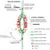
Figure 7. Cartoon depicting the ependyma as a stem cell niche organized in midline and lateral spatial domains
The heterogeneous molecular and functional phenotypes are color coded or illustrated with representative data. Some of our working hypotheses brought about by our current findings are indicated with interrogation marks.

Similar articles

Cited by

References

    1. Ming GL, Song H. Adult neurogenesis in the mammalian central nervous system. Annu Rev Neurosci. 2005;28:223–250. - PubMed
    1. Lledo PM, Alonso M, Grubb MS. Adult neurogenesis and functional plasticity in neuronal circuits. Nat Rev Neurosci. 2006;7:179–193. - PubMed
    1. Hugnot JP, Franzen R. The spinal cord ependymal region: a stem cell niche in the caudal central nervous system. Front Biosci. 2011;16:1044–1059. - PubMed
    1. Meletis K, Barnabé-Heider F, Carlén M, et al. Spinal cord injury reveals multilineage differentiation of ependymal cells. PLoS Biol. 2008;6:1494–1507. - PMC - PubMed
    1. Petit A, Sanders AD, Kennedy TE, et al. Adult spinal cord radial glia display a unique progenitor phenotype. PLoS One. 2011;6:e24538. - PMC - PubMed

Publication types