

Review
doi: 10.1002/adma.201201762.
Epub 2012 Jul 23.
Building bridges: leveraging interdisciplinary collaborations in the development of biomaterials to meet clinical needs
Affiliations
- PMID: 22821772
- PMCID: PMC3706713
- DOI: 10.1002/adma.201201762
Item in Clipboard
Review
Building bridges: leveraging interdisciplinary collaborations in the development of biomaterials to meet clinical needs
Adv Mater.
.
Abstract
Our laboratory at Rice University has forged numerous collaborations with clinicians and basic scientists over the years to advance the development of novel biomaterials and the modification of existing materials to meet clinical needs. This review highlights collaborative advances in biomaterials research from our laboratory in the areas of scaffold development, drug delivery, and gene therapy, especially as related to applications in bone and cartilage tissue engineering.
Copyright © 2012 WILEY-VCH Verlag GmbH & Co. KGaA, Weinheim.
Figures
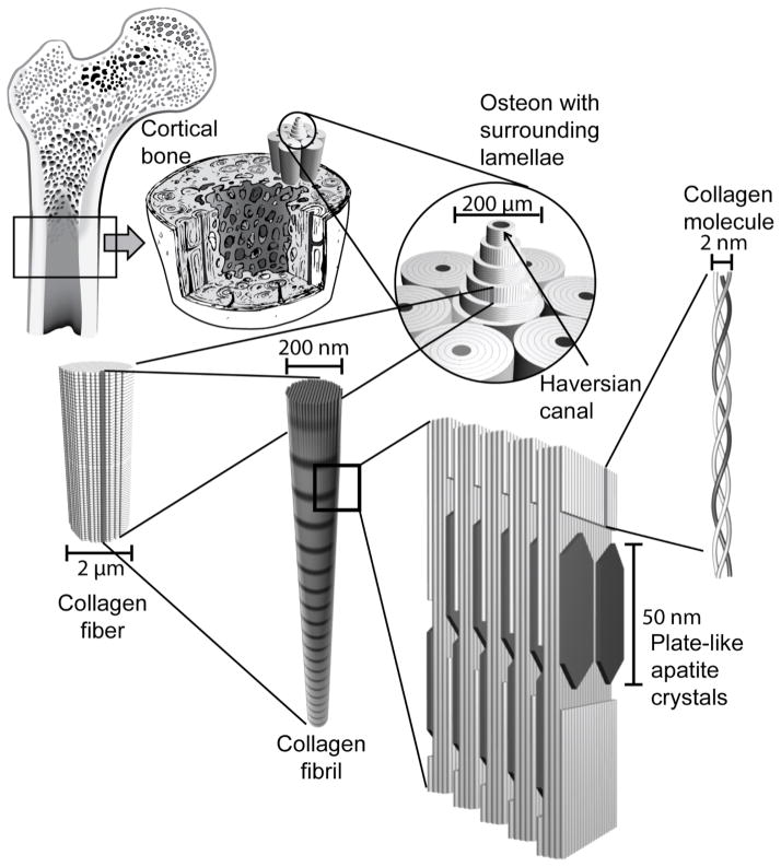
The hierarchical structure of bone. Cortical bone is composed of densely packed osteons made up of lamellae of collagen fibers surrounding a central Haversian canal. Collagen fibers are composed of bundles of collagen molecules called collagen fibrils. Plate-like hydroxyapatite crystals are deposited in the gaps of the collagen molecule structures within collagen fibrils.
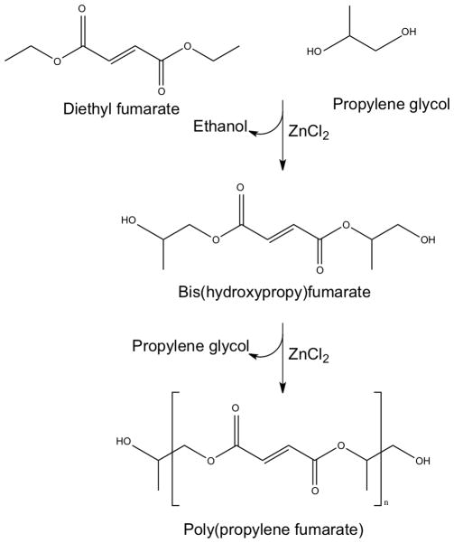
Two-step synthesis of poly(propylene fumarate) from diethyl fumarate and propylene glycol catalyzed by ZnCl2. Reproduced with permission [17].
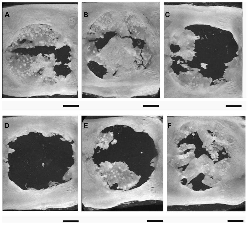
MicroCT generated maximum intensity projections of rat cranial defects 12 weeks after implantation of various poly(propylene fumarate)/gelatin microparticle (GMP) composite scaffolds. (A) Scaffold loaded with 1.25 mg BMP-2-loaded 40 mM basic GMP demonstrated 28.5% bone fill. (B) Scaffold loaded with 1.25 mg BMP-2-loaded 40 mM basic GMPs and 1.25 mg VEGF-loaded 10 mM acidic GMPs demonstrated 40% bone fill. (C) Scaffold loaded with 1.25 mg BMP-2-loaded 40 mM basic GMP and 2.5 mg VEGF-loaded 10 mM acidic GMP demonstrated 10.9% bone fill. (D) Scaffold loaded with 0.63 mg BMP-2-loaded 40 mM basic GMP demonstrated 3.1% bone fill. (E) Scaffold loaded with 0.63 mg BMP-2-loaded 40 mM basic GMP and 1.25 mg VEGF-loaded 10 mM acidic GMP demonstrated 15.1% bone fill. (F) Scaffold loaded with 0.63 mg BMP-2-loaded 40 mM basic GMP and 2.5 mg VEGF-loaded 10 mM acidic GMP demonstrated 35% bone fill. Bars represent 2 mm. Reproduced with permission from [36].
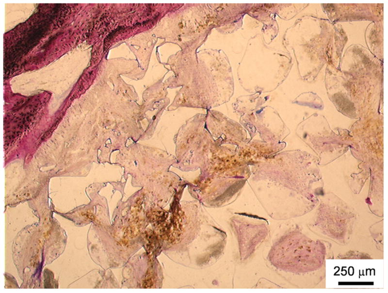
Histological section of a poly(propylene fumarate) scaffold stained using methylene blue and basic fuchsin (which stains nuclei purple, collagen and connective tissue blue, and cytoplasm and smooth muscle cells pink) 12 weeks after implantation into a goat femoral condyle. The top left area demonstates the in vivo breakdown of PPF into smaller fragments as well as soft tissue infiltration with minimal inflammation. Reproduced with permission from [39].
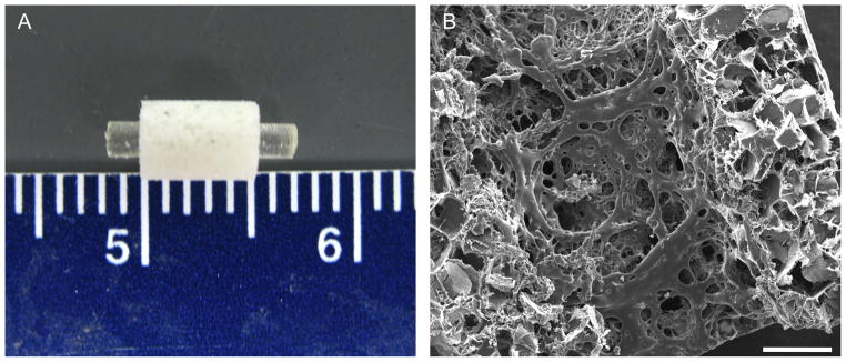
Combination scaffolds composed of a solid intramedullary poly(propylene fumarate) rod surrounded by a porous poly(propylene fumarate) sleeve to be used in a rat segmental femoral defect shown grossly (A) and microscopically through scanning electron microscopy (B). Scale bar in (B) represents 500 μm. Reproduced with permission from [48].
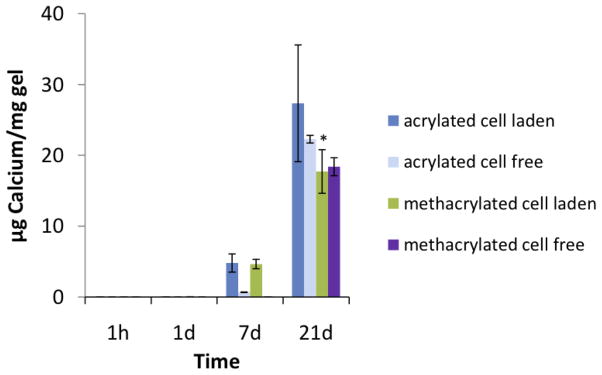
Calcium content of wet mesenchymal stem cell-laden and cell-free poly(N-isopropyl acrylamide)-based hydrogels containing pentaerythritol diacrylate monstearate hydrophobic domains after culture in osteogenic medium (n = 3–5). Calcium content was undetectable at the 1h and 1d time points. Reproduced with permission from [59].
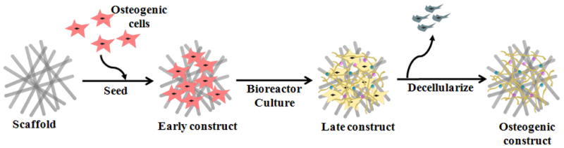
Generation of an extracellular matrix-scaffold construct for bone regeneration. A naked scaffold is first seeded with osteogenic progenitor cells. The cell/scaffold construct is then cultured in a bioreactor under flow perfusion conditions, where cells lay down extracellular matrix that coats the scaffold. By decellularizing the construct, an extracellular matrix-coated scaffold capable of supporting osteogenic differentiation of progenitor cells is obtained. Reproduced with permission from [171].
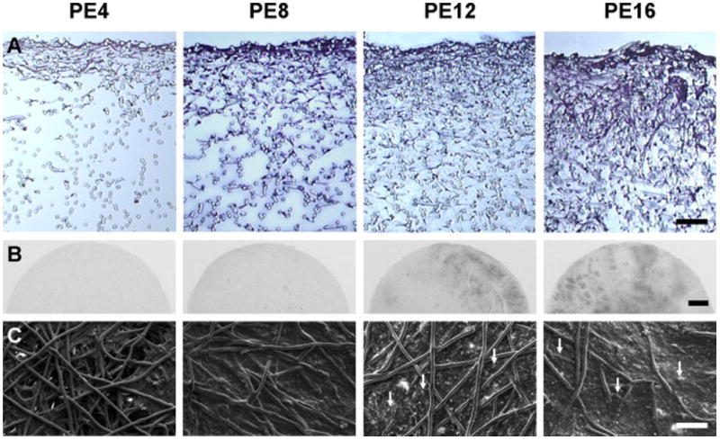
MSCs were cultured on poly(ε-caprolactone) microfiber scaffolds under flow perfusion conditions in a bioreactor to examine the effect of culture duration on mineralized extracellular matrix deposition. PE 4, PE 8, PE 12 and PE 16 represent the PCL/extracellular matrix (PE) constructs that were generated in flow perfusion culture of increasing durations (4, 8, 12 and 16 days, respectively). Flow perfusion conditions augmented the distribution of cells and extracellular matrix proteins over time, as observed via histological sections stained with hematoxylin and eosin, as shown in (A). Scale bar represents 100 μm. X-ray imaging indicated that radio-opaque regions of mineralized matrix increased over time, as shown in (B). Scale bar represents 1 mm. Scanning electron microscopy was used to visualize the surface morphology of constructs, as shown in (C). Arrows indicate mineral nodules and scale bar represents 100 μm. Reproduced with permission from [71].
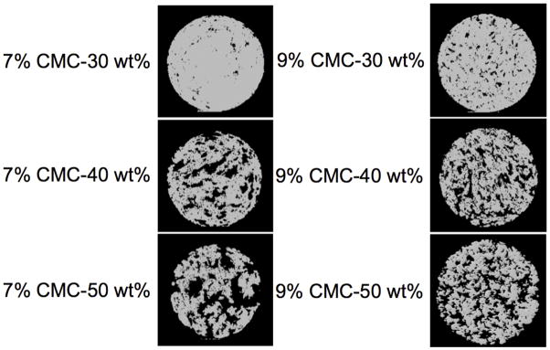
MicroCT images of cross-sections of cylindrical poly(methyl methacrylate) implants (10 mm diameter × 6 mm height) of varying porosity. Either 7 or 9 wt% carboxymethylcellulose hydrogels were incorporated at 30, 40, or 50 wt% with poly(methyl methacrylate) cement to form the above porous implants. Digital cross sections of the implants were made by slicing through the center of the axially oriented implant. Reproduced with permission from [85].
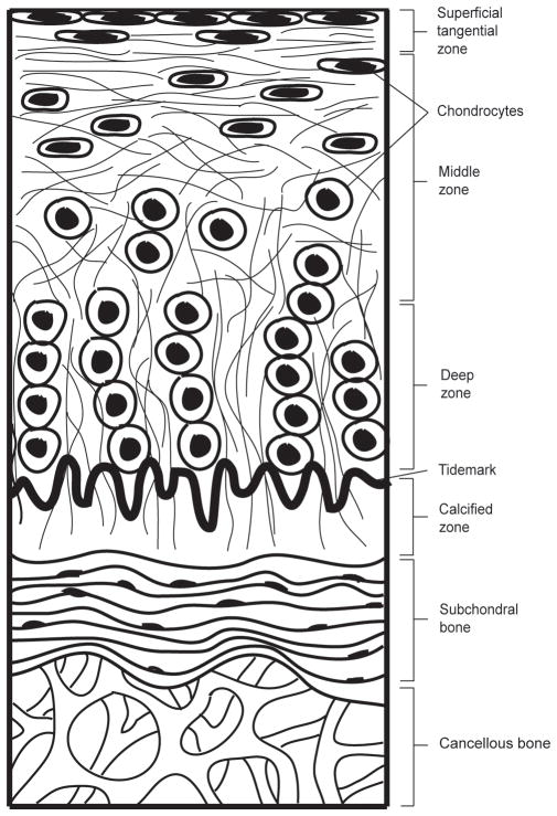
Structure of articular cartilage. The articular cartilage is divided into four distinct zones – superficial tangential zone, middle zone, deep zone and calcified zone. Within each zone, chondrocytes and collagen fibers are uniquely organized. The underlying subchondral bone and cancellous bone provide support to the articular cartilage layer.
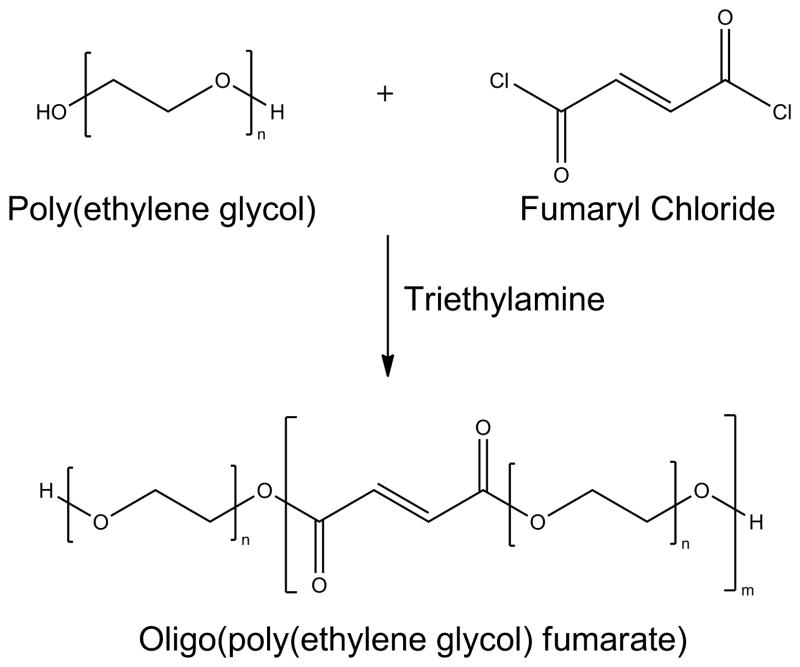
The oligo(poly(ethylene glycol) fumarate) macromer is synthesized by reacting poly(ethylene glycol) with fumaryl chloride in the presence of triethylene amine.
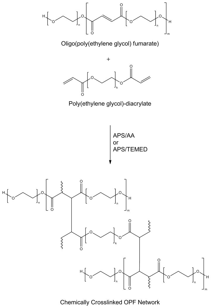
Poly(ethylene glycol)-diacrylate can be used to cross-link oligo(poly(ethylene glycol) fumarate) in the presence of ammonium persulfate/ascorbic acid (APS/AA) or ammonium persulfate/tetramethylethylenediamine (APS/TEMED) to form a hydrogel.
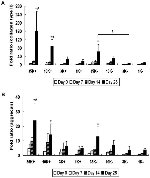
Collagen type II and aggregan gene expression over time. Poly(ethylene glycol) of four different molecular weights (35000 g mol−1, 10000 g mol−1, 3300 g mol−1 and 1000 g mol−1) were used to prepare oligo(poly(ethylene glycol) fumarate) macromers of four repeating units (OPF 35K, OPF 10K, OPF 3K and OPF 1K, respectively). (A) depicts collagen type II gene expression and (B) depicts aggrecan gene expression for OPF 35K, 10K, 3K and 1K hydrogel composites encapsulating rabbit MSCs and TGF-β1-loaded microparticles (+) or rabbit MSCs and blank microparticles (−). Results are presented as a fold ratio after being normalized to GAPDH values. The OPF 35K- group shows the average expression level of controls (Day 0), represented as one. (*) indicates that within a given hydrogel formulation, a significantly higher (p < 0.05) gene expression than the Day 0 value (control) was observed. (#) indicates samples which had significantly higher (p < 0.05) gene expression than other OPF formulations at the same time point. Error bars represent means ± standard deviation for n=4. Reproduced with permission from [121].
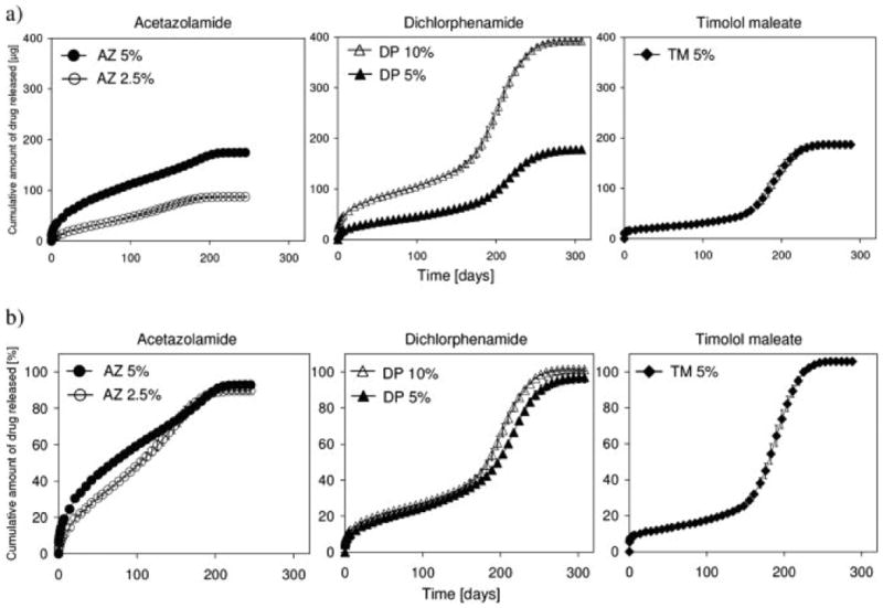
Cumulative amount of model ophthalmic drug release over time from photo-cross-linked poly(propylene fumarate)/poly(N-vinyl pyrrolidone) (PPF/PVP) (3:2 ratio) matrices in phosphate buffered saline at 37°C measured (a) absolutely (μg) and (b) relatively (%). The drugs acetazolamide (AZ) (2.5 and 5 wt%), dichlorphenamide (DP) (5 and 10 wt%), and timolol maleate (TM) (5 wt%) were incorporated into the PPF/PVP matricies. Data represent means ± standard deviation for n = 3. Reproduced with permission from [156].
References
-
- Myeroff C, Archdeacon M. J Bone Joint Surg Am. 2011;93:2227. - PubMed
-
- Sawin PD, Traynelis VC, Menezes aH. J Neurosurg. 1998;88:255. - PubMed
-
- Laurencin C, Khan Y, El-Amin SF. Expert Rev Med Devices. 2006;3:49. - PubMed
-
- Greenwald aS, Boden SD, Goldberg VM, Khan Y, Laurencin CT, Rosier RN. J Bone Joint Surg Am. 2001;83-A(Suppl):98. - PubMed
-
- Polak JM, Bishop AE. Ann N Y Acad Sci. 2006;1068:352. - PubMed
Publication types
MeSH terms
Substances
Grants and funding
LinkOut - more resources
Full Text Sources
