Review
. 2012 Jul 20;111(3):375-85.
doi: 10.1161/CIRCRESAHA.110.223842.

Understanding cardiomyopathy phenotypes based on the functional impact of mutations in the myosin motor

Affiliations
Review

Understanding cardiomyopathy phenotypes based on the functional impact of mutations in the myosin motor

Jeffrey R Moore et al. Circ Res. .

Abstract

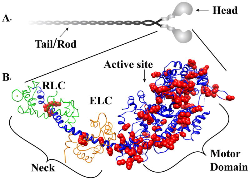
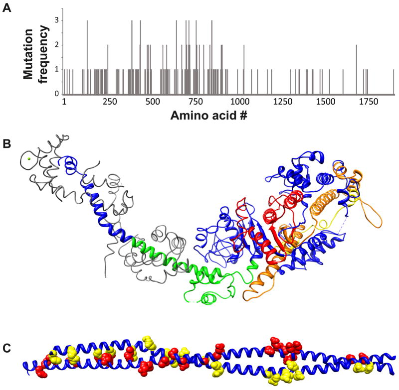
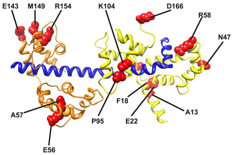
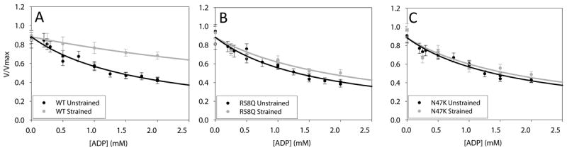
Hypertrophic (HCM) and dilated (DCM) cardiomyopathies are inherited diseases with a high incidence of death due to electric abnormalities or outflow tract obstruction. In many of the families afflicted with either disease, causative mutations have been identified in various sarcomeric proteins. In this review, we focus on mutations in the cardiac muscle molecular motor, myosin, and its associated light chains. Despite the >300 identified mutations, there is still no clear understanding of how these mutations within the same myosin molecule can lead to the dramatically different clinical phenotypes associated with HCM and DCM. Localizing mutations within myosin's molecular structure provides insight into the potential consequence of these perturbations to key functional domains of the motor. Review of biochemical and biophysical data that characterize the functional capacities of these mutant myosins suggests that mutant myosins with enhanced contractility lead to HCM, whereas those displaying reduced contractility lead to DCM. With gain and loss of function potentially being the primary consequence of a specific mutation, how these functional changes trigger the hypertrophic response and lead to the distinct HCM and DCM phenotypes will be the future investigative challenge.


Figures

Figure. 1
Figure. 1. Structure the myosin molecule
Panel A Myosin is a hexameric protein consisting of two myosin heavy chain polypeptides that self-associate via an alpha helical coild-coil rod or tail domain. Each globular head domain is associated with two light chains one Essentail light chain (ELC) and one Regulatory light chain (RLC). Panel B: The atomic structure of the myosin head domain is shown. The heavy chain is shown in blue. The essential light chain and regulatory light chain are colored in grey. Small conformational changes in the motor domain are amplified by the light chain binding neck domain to produce movement and force. Cardiomyopathy-linked mutations (highlighted in red) are distributed throughout the head domain.
Figure 2
Figure 2. HCM and DCM mutations are distributed throughout the molecule
Panel A: Distribution of mutations along the myosin heavy chain sequence. Mutations cluster in several regions: The ATP binding region (~130-260), the highly conserved regions implicated in actin binding (385-515 and 577-611), the long helix that extends from the SH1 and SH2 helix through the converter region and into the ELC binding domain (~705-810) and the S2 region at the junction between the motor domain and the filament forming rod domain. Panel B: Cardiomyopathy mutation clusters mapped on the atomic structure of chicken fast skeletal muscle myosin. ATP binding region (red). Actin binding region (yellow and orange). Converter/ELC binding (green). Panel C: Mutations in the S2 region mutations on one chain of the dimer are colored red while mutations in the other chain are highlight in yellow.
Figure. 3
Figure. 3. HCM mutations (red) mapped onto the chicken skeletal myosin light chain structures
The C-terminal region of the MyHC neck is shown in blue, the RLC in green and the ELC in orange.
Figure. 4
Figure. 4. HCM mutations in the myosin RLC disrupt myosin strain sensitive biochemistry
Porcine cardiac myosin velocity as a function of ADP. (gray) loaded (black) unloaded. Panels A, B and C are Wild-type, R58Q and N47K respectively.
Figure. 5
Figure. 5. Cardiomyopathy mutations in the myosin rod
Panels A and B: Mutation location corresponding to the heptad repeat (abcdefg)n are shown in purple, blue, green, cyan, yellow, red, and pink respectively. Panel A open circles; Panel B filled circles Panel C: helical wheel representation of the canonical coiled-coil structure. Hydrophobic interactions between the a and d postions and salt bridges between the e and g positions stabilize the structure.
Figure 6
Figure 6. Thick filament myosin interacting head motif
Panel A: The interacting head motif docked onto the electron density map of the cardiac muscle thick filament. The interacting head motif contains interactions between the heads of the dimer and additional interactions between the heads and the thick filament backbone. In the motif, one head has its actin binding domain “free” to potentially interact with actin while the other head is in a “blocked” configuration where its actin binding domain interacts with the converter region of the free head. Panel B: Several cardiomyopathy mutations map to regions that could potentially alter the interacting head motif. Potentially interacting residues are highlighted as examples (red spheres).

References

    1. Olivotto I, Cecchi F, Poggesi C, Yacoub MH. Developmental origins of hypertrophic cardiomyopathy phenotypes: A unifying hypothesis. Nat Rev Cardiol. 2009;6:317–321. - PubMed
    1. Liew CC, Dzau VJ. Molecular genetics and genomics of heart failure. Nat Rev Genet. 2004;5:811–825. - PubMed
    1. Marian AJ, Roberts R. Molecular basis of hypertrophic and dilated cardiomyopathy. Tex Heart Inst J. 1994;21:6–15. - PMC - PubMed
    1. Ramaraj R. Hypertrophic cardiomyopathy: Etiology, diagnosis, and treatment. Cardiol Rev. 2008;16:172–180. - PubMed
    1. Kamisago M, Sharma SD, DePalma SR, Solomon S, Sharma P, McDonough B, Smoot L, Mullen MP, Woolf PK, Wigle ED, Seidman JG, Seidman CE. Mutations in sarcomere protein genes as a cause of dilated cardiomyopathy. N Engl J Med. 2000;343:1688–1696. - PubMed

Publication types