. 2012 Sep 15;303(6):G675-85.
doi: 10.1152/ajpgi.00152.2012. Epub 2012 Jul 19.

Colonic mucosal microbiome differs from stool microbiome in cirrhosis and hepatic encephalopathy and is linked to cognition and inflammation

Affiliations

Colonic mucosal microbiome differs from stool microbiome in cirrhosis and hepatic encephalopathy and is linked to cognition and inflammation

Jasmohan S Bajaj et al. Am J Physiol Gastrointest Liver Physiol. .

Abstract

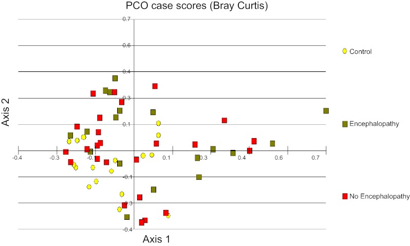
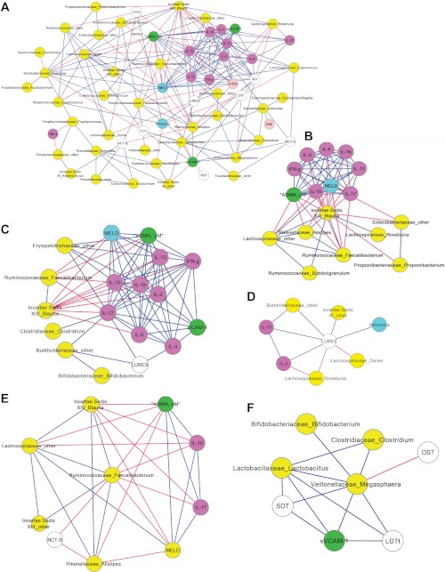
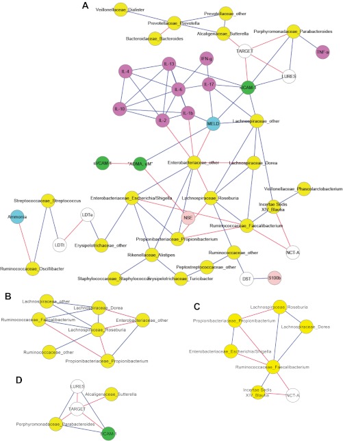
Although hepatic encephalopathy (HE) is linked to the gut microbiota, stool microbiome analysis has not found differences between HE and no-HE patients. This study aimed to compare sigmoid mucosal microbiome of cirrhotic patients to controls, between HE vs. no-HE patients, and to study their linkage with cognition and inflammation. Sixty cirrhotic patients (36 HE and 24 no-HE) underwent cognitive testing, stool collection, cytokine (Th1, Th2, Th17, and innate immunity), and endotoxin analysis. Thirty-six patients (19 HE and 17 no-HE) and 17 age-matched controls underwent sigmoid biopsies. Multitag pyrosequencing (including autochthonous genera, i.e., Blautia, Roseburia, Fecalibacterium, Dorea) was performed on stool and mucosa. Stool and mucosal microbiome differences within/between groups and correlation network analyses were performed. Controls had significantly higher autochthonous and lower pathogenic genera compared with cirrhotic patients, especially HE patients. HE patients had worse MELD (model for end-stage liver disease) score and cognition and higher IL-6 and endotoxin than no-HE. Mucosal microbiota was different from stool within both HE/no-HE groups. Between HE/no-HE patients, there was no difference in stool microbiota but mucosal microbiome was different with lower Roseburia and higher Enterococcus, Veillonella, Megasphaera, and Burkholderia abundance in HE. On network analysis, autochthonous genera (Blautia, Fecalibacterium, Roseburia, and Dorea) were associated with good cognition and decreased inflammation in both HE/no-HE, whereas genera overrepresented in HE (Enterococcus, Megasphaera, and Burkholderia) were linked to poor cognition and inflammation. Sigmoid mucosal microbiome differs significantly from stool microbiome in cirrhosis. Cirrhotic, especially HE, patients' mucosal microbiota is significantly different from controls with a lack of potentially beneficial autochthonous and overgrowth of potentially pathogenic genera, which are associated with poor cognition and inflammation.


Figures

Fig. 1.
Fig. 1.
Principal component analysis (PCO) of sigmoid mucosal microbiome between controls and patients with and without hepatic encephalopathy (HE). The first principal component accounts for 19% of the variance and the second component accounts for 17% of the variance for a total of 36% of the variance. We found significant clustering of the controls with each other. Additionally, the no-HE patients cluster around the controls whereas HE patients were scattered further from the central control cluster. Yellow: controls; red: no hepatic encephalopathy; green: hepatic encephalopathy patients.
Fig. 2.
Fig. 2.
Correlation network and subnetworks of the mucosal microbiome of HE patients. Connecting lines in red indicate a significant negative whereas those in blue mean a significantly positive correlation. Nodes in yellow are bacterial genera, purple are inflammatory cytokines, white are cognitive tests, blue are clinical variables, green are markers of endothelial activation, and light pink are neuroglial markers. A high score on digit symbol (DST) and targets indicates good cognition whereas a high score on the rest of the cognitive tests indicates poor performance. SDT, serial dotting; LDTt, line tracing test time; NCT-A/B, number connection test A/B. A: correlation network of the mucosal microbiome of HE patients. As can be seen, autochthonous genera belonging to the Ruminococcaceae, Lachnospiraceae, and Incertae Sedis families are associated with good cognition, lower model for end-stage liver disease (MELD) score, lower ammonia, and decreased inflammation. Subnetworks from this complex network are displayed in the following panels. B: subnetwork of the HE mucosa microbiome showing the negative correlation of the autochthonous bacteria to MELD score and inflammation. C: subnetwork of the HE mucosa microbiome showing the negative correlation of the inflammatory cytokines, particularly IL-17 with autochthonous bacteria and positive correlation with lures (indicating worse cognition with increased inflammation), endothelial activation [soluble intravascular adhesion molecule (sICAM-1)], MELD score, and nonautochthonous bacterial genera (Burkholderiaceae, Erysipelothricaceae). D: a high lure number indicates poor cognition. This subnetwork of the HE mucosa microbiome shows that lures are negatively correlated with autochthonous bacterial genera (Roseburia and Dorea) whereas they are correlated positively with Burkholderiaceae and Incertae Sedis XI and as expected with ammonia and inflammatory cytokines. E: a high number on NCT-B indicates poor performance. This subnetwork of the HE mucosal microbiome shows a negative correlation, i.e., good NCT-B performance with the abundances of Ruminococcaceae Fecalibacterium. This autochthonous genus has been associated with lower MELD score and lower inflammation (IL-17 and IL-10) and is positively correlated with other beneficial autochthonous bacteria. F: Megasphaera was significantly more abundant in HE; in this subnetwork Megasphaera abundance is significantly correlated with soluble vascular adhesion molecule (sVCAM-1) (marker of endothelial activation) and with poor cognitive performance (a high score on SDT and LDTt indicates poor whereas a high score on DST indicates good cognitive performance).
Fig. 3.
Fig. 3.
Correlation network and subnetworks of the mucosal microbiome of patients without HE. Connecting lines in red indicate a significant negative whereas those in blue mean a significantly positive correlation. Nodes in yellow are bacterial genera, purple are inflammatory cytokines, white are cognitive tests, blue are clinical variables, green are markers of endothelial activation, and light pink are neuroglial markers. A high score on DST and targets indicates good cognition whereas a high score on the rest of the cognitive tests indicates poor performance. A: correlation network of the mucosal microbiome of patients without HE. Autochthonous genera belonging to the Ruminococcaceae, Lachnospiraceae, and Incertae Sedis families are associated with good cognition and lower MELD, ammonia, and inflammation. B: this subnetwork of patients without HE shows that bacteria genera belonging to autochthonous families (Ruminococcaceae and Lachnospiraceae) are positively correlated with each other while negatively correlated with potentially pathogenic Enterobacteriaceae and Propionibacterium. C: this subnetwork of the no-HE mucosal microbiome again shows the positive correlation of the autochthonous bacteria with each other and a negative correlation with time required to complete NCT-A, which indicates good cognitive performance. D: a high score on targets and low score on lures indicates good cognitive performance. We again found a correlation between poor performance on lures and targets with genera belonging to Porphyromonadaceae and Alcaligenaceae.

References

    1. Ahern PP, Schiering C, Buonocore S, McGeachy MJ, Cua DJ, Maloy KJ, Powrie F. Interleukin-23 drives intestinal inflammation through direct activity on T cells. Immunity 33: 279–288, 2010 - PMC - PubMed
    1. Atarashi K, Tanoue T, Shima T, Imaoka A, Kuwahara T, Momose Y, Cheng G, Yamasaki S, Saito T, Ohba Y, Taniguchi T, Takeda K, Hori S, Ivanov II, Umesaki Y, Itoh K, Honda K. Induction of colonic regulatory T cells by indigenous Clostridium species. Science 331: 337–341, 2011 - PMC - PubMed
    1. Bajaj JS, Gillevet PM, Patel NR, Ahluwalia V, Ridlon JM, Kettenmann B, Schubert CM, Sikaroodi M, Heuman DM, Crossey MME, Bell DE, Hylemon PB, Fatouros PP, Taylor-Robinson SD. A longitudinal systems biology analysis of lactulose withdrawal in hepatic encephalopathy. Metab Brain Dis 27: 205–215, 2012 - PubMed
    1. Bajaj JS, Hafeezullah M, Franco J, Varma RR, Hoffmann RG, Knox JF, Hischke D, Hammeke TA, Pinkerton SD, Saeian K. Inhibitory control test for the diagnosis of minimal hepatic encephalopathy. Gastroenterology 135: 1591–1600, 2008 - PubMed
    1. Bajaj JS, Heuman DM, Wade JB, Gibson DP, Saeian K, Wegelin JA, Hafeezullah M, Bell DE, Sterling RK, Stravitz RT, Fuchs M, Luketic V, Sanyal AJ. Rifaximin improves driving simulator performance in a randomized trial of patients with minimal hepatic encephalopathy. Gastroenterology 140: 478–487, 2011 - PMC - PubMed

Publication types