. 2012 Sep;194(18):5080-90.
doi: 10.1128/JB.01092-12. Epub 2012 Jul 20.

Evidence for cyclic Di-GMP-mediated signaling in Bacillus subtilis

Affiliations

Evidence for cyclic Di-GMP-mediated signaling in Bacillus subtilis

Yun Chen et al. J Bacteriol. 2012 Sep.

Abstract

Cyclic di-GMP (c-di-GMP) is a second messenger that regulates diverse cellular processes in bacteria, including motility, biofilm formation, cell-cell signaling, and host colonization. Studies of c-di-GMP signaling have chiefly focused on Gram-negative bacteria. Here, we investigated c-di-GMP signaling in the Gram-positive bacterium Bacillus subtilis by constructing deletion mutations in genes predicted to be involved in the synthesis, breakdown, or response to the second messenger. We found that a putative c-di-GMP-degrading phosphodiesterase, YuxH, and a putative c-di-GMP receptor, YpfA, had strong influences on motility and that these effects depended on sequences similar to canonical EAL and RxxxR-D/NxSxxG motifs, respectively. Evidence indicates that YpfA inhibits motility by interacting with the flagellar motor protein MotA and that yuxH is under the negative control of the master regulator Spo0A∼P. Based on these findings, we propose that YpfA inhibits motility in response to rising levels of c-di-GMP during entry into stationary phase due to the downregulation of yuxH by Spo0A∼P. We also present evidence that YpfA has a mild influence on biofilm formation. In toto, our results demonstrate the existence of a functional c-di-GMP signaling system in B. subtilis that directly inhibits motility and directly or indirectly influences biofilm formation.


Figures

Fig 1
Fig 1
Genes predicted to be involved in c-di-GMP signaling in B. subtilis. (A) Schematic representation of c-di-GMP-mediated regulation of motility and biofilm formation in bacteria. (B) Domain composition and organization of eight putative c-di-GMP signaling proteins in B. subtilis, including four GGDEF domain proteins, two EAL domain proteins, one protein with both a GGDEF and an EAL domain, and a PilZ domain protein. GGDEF domains are shown in green, and EAL domains are shown in red. The predicted PilZ domain in YpfA is shown in purple. Additional domains predicted by Pfam are shown in white, and their annotations are as follows: 5TM-5TMR LYT, transmembrane region of the 5TM-LYT (5-transmembrane receptors of the LytS-YhcK type); DHHA1, DHH subfamily 1 members; MHYT, N-terminal triplet tandem repeat in bacterial signaling proteins; PAS, functions as a signal sensor; HDOD, HD/PDEase superfamily.
Fig 2
Fig 2
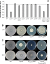
EAL domain protein YuxH and PilZ domain protein YpfA regulate swarming motility. (A) Assays of swarming motility by various single and triple mutants of putative c-di-GMP signaling genes. The sizes of the swarming zones on the plates inoculated with the mutants were normalized against that of the wild type. The results showed that the mutant of YuxH for a putative phosphodiesterase (CY9) and the triple mutant of YuxH YkoW YkuI (CY25) were severely impaired for swarming motility. All values are the averages of three replicates. (B) The severe defect in swarming motility by the yuxH mutant was substantially rescued by a second mutation in ypfA. Swarming zones of the wild type, ΔypfA (CY3), ΔyuxH (CY9), and ΔyuxH ΔypfA double mutant (CY30) were visualized after incubation of the swarming plates at 37°C for 5 h and at room temperature for another 12 h (see Materials and Methods). (C) YpfA overexpression had a strong, negative effect on swarming motility. No swarming motility was observed after 5 h of incubation in the ypfA-overexpressing strain (CY84). The inhibitory effect was even greater in a yuxH mutant strain overexpressing YpfA (CY85), in that the strain did not initiate swarming even at 20 h postinoculation.
Fig 3
Fig 3
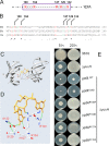
Conserved residues in the EAL domain of YuxH are critical for its activity, and yuxH is negatively regulated by Spo0A∼P. (A) Partial sequence alignment of YuxH of B. subtilis with homologs of YuxH from C. difficile and V. cholerae. Conserved EAL-like motifs are highlighted in blue and red. The sequence alignment was created using Clustal X 2.0 (56). (B) Swarming plates inoculated with the wild type, the yuxH mutant (CY9), or the yuxH mutant complemented with wild-type yuxH at amyE (CY87) or with a mutant allele of yuxH (88EIL90>88EDA90) at amyE (CY89). (C) The regulatory region of yuxH contains two imperfect Spo0A∼P binding sites (TTAGACA and TTCGTCA [highlighted in red]), which overlap the putative −35 region of the σA-dependent promoter (putative −35 and −10 regions are underlined). (D) Expression levels of the PyuxH-lacZ fusion in the wild type (blue diamonds; CY110) and the spo0A mutant (red squares; CY121) were determined based on β-galactosidase activities in DS medium. T0 represents start of stationary growth of cells.
Fig 4
Fig 4
Conserved PilZ domain residues in YpfA are critical for regulating motility. (A) Schematic showing conserved amino acid residues in the putative PilZ domain in YpfA. Conserved residues of the PilZ domain in YpfA, the putative c-di-GMP binding sites, are highlighted in red, and on the top their positions in the protein are marked. (B) Partial sequence alignment among YpfA of B. subtilis, PlzD of V. cholerae, and YcgR of E coli. Conserved residues in all three PilZ domains are highlighted in red. (C) The X-ray crystal structure of PlzD of V. cholerae complexed with c-di-GMP (4). The PlzD protein is shown in light gray, and the c-di-GMP molecule is shown in yellow. (D) Predicted interactions between c-di-GMP and conserved residues in PlzD. (E) The swarming phenotype of a yuxH mutant that overproduced YpfA, bearing the indicated mutations in the conserved residues in the PilZ domain.
Fig 5
Fig 5
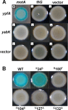
YpfA interacts with MotA for motility inhibition. (A) Bacterial two-hybrid assays were applied to test the interactions between YpfA and the putative targets MotA and FliG. YabK and vector only were used as controls. Five-microliter culture aliquots of E. coli BTH101 cells harboring derivatives of pKNT25 and pCH363 with cloned genes were spotted on plates supplemented with X-Gal, and colonies were visualized and imaged after 48 h of incubation at 23°C. A blue colony indicates strong interactions between the two fusion proteins, while a white colony implies no interactions. (B) The bacterial two-hybrid assays showed that mutations in the putative c-di-GMP binding sites in YpfA blocked interactions between YpfA and MotA. YpfAK24D was used as a control and was shown previously to be fully functional.
Fig 6
Fig 6
Effects of mutations in c-di-GMP signaling genes on biofilm colony morphology and pellicle formation. For biofilm colony development, individual colonies of the indicated mutants were grown on MSgg agar plates for 3 days at 23°C before imaging. Pellicles were formed in 6-well microtiter plates for 3 days at 23°C before imaging, with each well filled with 9 ml of MSgg liquid medium and inoculated with the indicated mutant cells.
Fig 7
Fig 7
The ypfA mutant forms robust central wrinkles in biofilm colonies. (A) Colony morphology of the ΔypfA mutant (CY3) and the wild type (3610) on MSgg agar plates was recorded every 24 h over a period of 72 h. (B) Details of the central parts of the biofilm colonies formed by the wild type and the ypfA mutant (CY3) on MSgg agar plates 3 days after inoculation were captured by using Zeiss Lumar stereomicroscope. Bars, in panels from left to right, represent 500, 300, 100, and 50 μm, respectively. (C) Expression levels of the two biofilm matrix operons (epsA-O and tapA) in the wild type and the ypfA mutant as measured by β-galactosidase activities. Reporter strains were grown to an OD600 of 1.0 in MSgg broth and assayed for β-galactosidase activities. Strains used in the assays were YC110 and CY437 for the wild type and the ypfA mutant bearing PepsA-lacZ and strains YC121 and CY438 for the wild type and the mutant bearing PtapA-lacZ. (D) YpfA acts upstream of KinA and KinB in regulating central winkle formation of the colony. Shown are colony morphologies of kinA kinB (RL4573), kinC kinD (RL5273), kinA kinB ypfA (CY230), and kinC kinD ypfA (CY231) on MSgg agar plates after 3 days of incubation at 23°C.
Fig 8
Fig 8
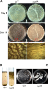
The ypfA mutation delays biofilm disassembly. (A) Pellicles formed by the wild type (3610) and the ypfA mutant (CY3) in MSgg liquid medium at day 4 (top panels) and day 14 (middle panels) postinoculation at 23°C. Details of the day 14 pellicles (lower panels) were also examined by using a stereomicroscope at ×12 magnification. (B) Day 4 pellicles formed by the wild type or the ypfA mutant cells were subjected to vortexing. The ypfA mutant pellicles remained as large pieces and floated to the air-liquid interface in the test tube after vortexing, whereas the wild-type pellicles broke into smaller pieces and sunk to the bottom of the test tube after vortex treatment. (C) Similar to the results shown in panel B, pellicles formed by the wild-type and the ypfA mutant cells after vortex treatment were compared in petri dishes.

Similar articles

Cited by

References

    1. Amikam D, Galperin M. 2006. PilZ domain is part of the bacterial c-di-GMP binding protein. Bioinformatics 22:3–9 - PubMed
    1. Antoniewski C, Savelli B, Stragier P. 1990. The spoIIJ gene, which regulates early developmental steps in Bacillus subtilis, belongs to a class of environmentally responsive genes. J. Bacteriol. 172:86–93 - PMC - PubMed
    1. Ausmees N, et al. 2001. Genetic data indicate that proteins containing the GGDEF domain possess diguanylate cyclase activity. FEMS Microbiol. lett. 204:163–167 - PubMed
    1. Benach J, et al. 2007. The structural basis of cyclic diguanylate signal transduction by PilZ domains. EMBO J. 26:5153–5166 - PMC - PubMed
    1. Beyhan S, Odell LS, Yildiz FH. 2008. Identification and characterization of cyclic diguanylate signaling systems controlling rugosity in Vibrio cholerae. J. Bacteriol. 190:7392–7405 - PMC - PubMed

Publication types

Substances

LinkOut - more resources