. 2012 Sep;194(18):5110-7.
doi: 10.1128/JB.00958-12. Epub 2012 Jul 20.

The primosomal protein DnaD inhibits cooperative DNA binding by the replication initiator DnaA in Bacillus subtilis

Affiliations

The primosomal protein DnaD inhibits cooperative DNA binding by the replication initiator DnaA in Bacillus subtilis

Carla Y Bonilla et al. J Bacteriol. 2012 Sep.

Abstract

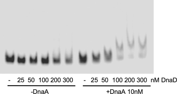
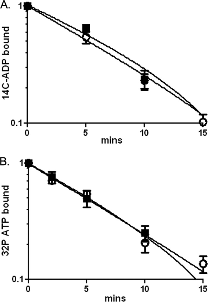
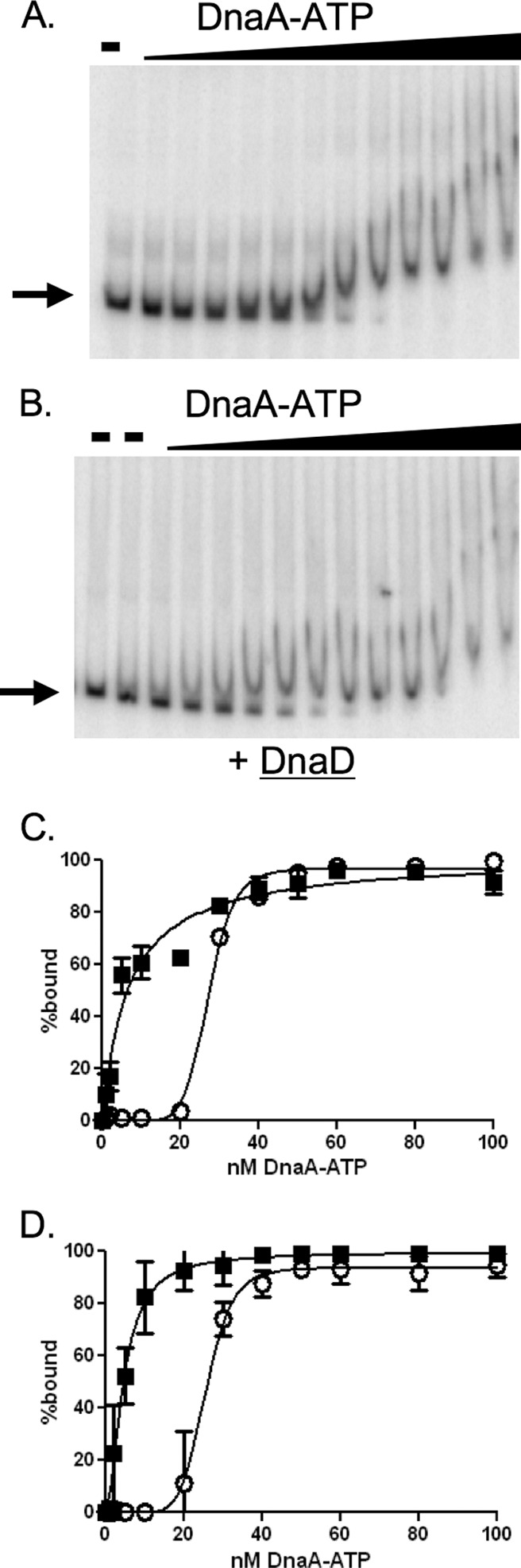
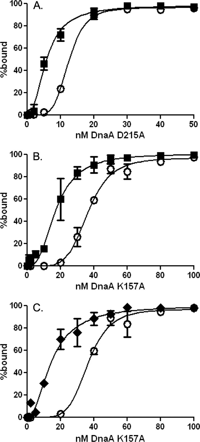
DnaA is an AAA+ ATPase and the conserved replication initiator in bacteria. Bacteria control the timing of replication initiation by regulating the activity of DnaA. DnaA binds to multiple sites in the origin of replication (oriC) and is required for recruitment of proteins needed to load the replicative helicase. DnaA also binds to other chromosomal regions and functions as a transcription factor at some of these sites. Bacillus subtilis DnaD is needed during replication initiation for assembly of the replicative helicase at oriC and during replication restart at stalled replication forks. DnaD associates with DnaA at oriC and at other chromosomal regions bound by DnaA. Using purified proteins, we found that DnaD inhibited the ability of DnaA to bind cooperatively to DNA and caused a decrease in the apparent dissociation constant. These effects of DnaD were independent of the ability of DnaA to bind or hydrolyze ATP. Other proteins known to regulate B. subtilis DnaA also affect DNA binding, whereas much of the regulation of Escherichia coli DnaA affects nucleotide hydrolysis or exchange. We found that the rate of nucleotide exchange for B. subtilis DnaA was high and not affected by DnaD. The rapid exchange is similar to that of Staphylococcus aureus DnaA and in contrast to the low exchange rate of Escherichia coli DnaA. We suggest that organisms in which DnaA has a high rate of nucleotide exchange predominantly regulate the DNA binding activity of DnaA and that those with low rates of exchange regulate hydrolysis and exchange.


Figures

Fig 1
Fig 1
Effects of different concentrations of DnaD on DnaA binding to oriC. Representative gel of the radiolabeled DNA probe from the oriC region with different amounts of DnaD-His, in the absence of DnaA (left six lanes) or the presence of 10 nM DnaA-ATP (right six lanes). Concentrations of DnaD-His are 0 (−), 25, 50, 100, 200, or 300 nM and are indicated below each lane.
Fig 2
Fig 2
DnaD does not affect nucleotide exchange by DnaA. The amount of [14C]ADP (A) or [32P]ATP (B) bound to DnaA (300 nM) at various times after addition of unlabeled ATP (2 mM) at 37°C was measured by filter binding in the absence (open circles) and presence (filled squares) of DnaD-His6 (600 nM). Data are averages of triplicates ± standard errors and are normalized to the starting amount of radioactivity in the absence of unlabeled ATP.
Fig 3
Fig 3
DnaD inhibits cooperative binding of DnaA to DNA. Representative gels and binding curves measuring binding of DnaA-ATP to DNA (50 pM) with and without purified DnaD-His6 (300 nM) are shown. DnaA concentrations used were 0, 1, 2, 5, 10, 20, 30, 40, 50, 60, 80, 100, and 200 nM. (A and B) Representative gels with increasing concentrations of DnaA-ATP incubated with template DNA from the oriC region in the absence (A) or presence (B) of DnaD-His6. Probe with no added protein is shown in the first lane of both panels. Probe with DnaD-His6 and no DnaA is shown in the second lane of panel B. (C and D) Data from three independent gel shift assays using template DNA from the oriC region (C) or the yydA region (D) are plotted as percent DNA bound versus the concentration of DnaA-ATP, in the absence (open circles) and presence (filled squares) of DnaD-His6. In experiments with the DNA fragment from the oriC region (C), the calculated Hill coefficient for DnaA-ATP was 8.6 in the absence of DnaD-His6 and 1 in the presence of DnaD-His6. The apparent Kd for DnaA-ATP was 27 nM in the absence and 6.6 nM in the presence of DnaD-His6. In experiments with the DNA fragment from the yydA region (D), the calculated Hill coefficient for DnaA-ATP was 6 in the absence of DnaD-His6 and 1 in the presence of DnaD-His6. The apparent Kd for DnaA-ATP was 25 nM in the absence and 5 nM in the presence of DnaD-His6.
Fig 4
Fig 4
Effects of DnaD and YabA on DnaA binding to DNA are independent of ATPase activity. Binding curves of DnaA mutants defective in ATP hydrolysis, DnaA(D215A)-ATP (A), and ATP binding, DnaA(K157A) (B and C), within the DNA fragment from the oriC region. DnaA concentrations tested were 0, 1, 2, 5, 10, 20, 30, 40, 50, 60, 80, 100, and 200 nM. (A and B) Binding in the absence (open circles) and presence (filled squares) of DnaD-His6 (300 nM). (C) Binding in the absence (open circles) and presence (filled diamonds) of His6-YabA (700 nM). For the DnaA mutant defective in ATP binding, DnaA(K157A), the presence of YabA reduced the Hill coefficient from 5.6 to 2 and the apparent Kd from 36 nM to 14.7 nM.

References

    1. Atlung T, Clausen ES, Hansen FG. 1985. Autoregulation of the dnaA gene of Escherichia coli K12. Mol. Gen. Genet. 200:442–450 - PubMed
    1. Bramhill D, Kornberg A. 1988. Duplex opening by dnaA protein at novel sequences in initiation of replication at the origin of the E. coli chromosome. Cell 52:743–755 - PubMed
    1. Braun RE, O'Day K, Wright A. 1985. Autoregulation of the DNA replication gene dnaA in E. coli K-12. Cell 40:159–169 - PubMed
    1. Breier AM, Grossman AD. 2009. Dynamic association of the replication initiator and transcription factor DnaA with the Bacillus subtilis chromosome during replication stress. J. Bacteriol. 191:486–493 - PMC - PubMed
    1. Bruand C, Ehrlich SD, Janniere L. 1995. Primosome assembly site in Bacillus subtilis. EMBO J. 14:2642–2650 - PMC - PubMed

Publication types

MeSH terms

LinkOut - more resources