. 2012 Sep;194(18):5101-9.
doi: 10.1128/JB.00932-12. Epub 2012 Jul 20.

Basis for the essentiality of H-NS family members in Pseudomonas aeruginosa

Affiliations

Basis for the essentiality of H-NS family members in Pseudomonas aeruginosa

Sandra Castang et al. J Bacteriol. 2012 Sep.

Abstract

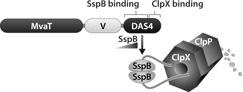
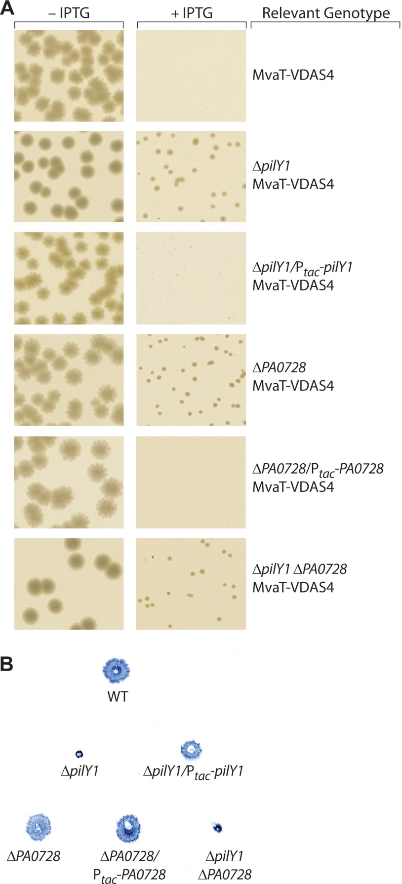
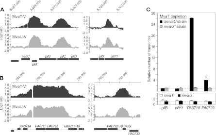
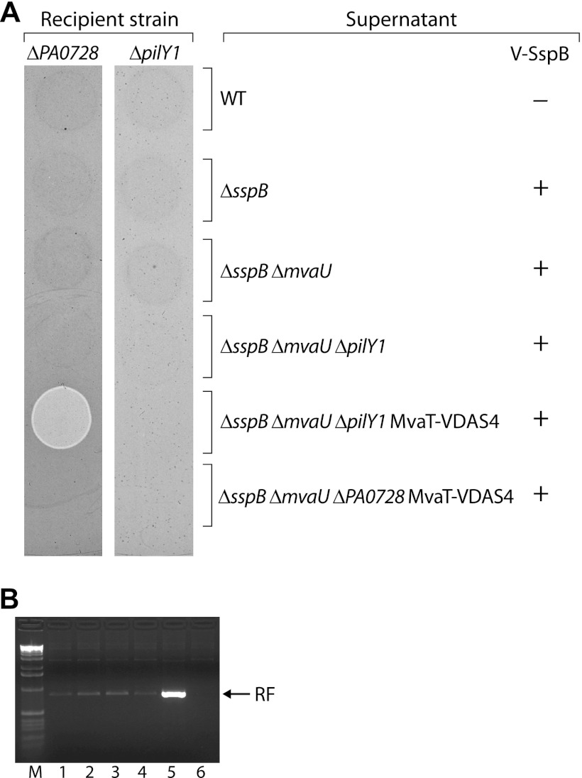
Members of the histone-like nucleoid-structuring (H-NS) family of proteins have been shown to play important roles in silencing gene expression and in nucleoid compaction. In Pseudomonas aeruginosa, the two H-NS family members MvaT and MvaU are thought to bind the same AT-rich regions of the chromosome and function coordinately to control a common set of genes. Here we present evidence that the loss of both MvaT and MvaU cannot be tolerated because it results in the production of Pf4 phage that superinfect and kill cells or inhibit their growth. Using a ClpXP-based protein depletion system in combination with transposon mutagenesis, we identify mutants of P. aeruginosa that can tolerate the depletion of MvaT in an ΔmvaU mutant background. Many of these mutants contain insertions in genes encoding components, assembly factors, or regulators of type IV pili or contain insertions in genes of the prophage Pf4. We demonstrate that cells that no longer produce type IV pili or that no longer produce the replicative form of the Pf4 genome can tolerate the loss of both MvaT and MvaU. Furthermore, we show that the loss of both MvaT and MvaU results in an increase in expression of Pf4 genes and that cells that cannot produce type IV pili are resistant to infection by Pf4 phage. Our findings suggest that type IV pili are the receptors for Pf4 phage and that the essential activities of MvaT and MvaU are to repress the expression of Pf4 genes.


Figures

Fig 1
Fig 1
Schematic representation of the ClpXP-based protein depletion system. The MvaT-VDAS4 protein is depicted. V corresponds to the VSV-G epitope tag. The DAS4 tag allows for depletion of MvaT-VDAS4 in a manner that is dependent upon the intracellular concentration of the adaptor protein SspB that feeds MvaT-VDAS4 to the ClpXP protease complex.
Fig 2
Fig 2
Deletion of pilY1 or PA0728 results in cells that can tolerate the loss of both MvaT and MvaU. (A) Colonies from cells of MvaT depletion strains carrying the plasmid pV-SspB that were grown overnight at 37°C on LB agar plates that either contained IPTG (+IPTG) or did not (−IPTG); images are from separate agar plates. All cells contained deletions of sspB and mvaU, and additional relevant genotypes are indicated. MvaT-VDAS4, PAO1 ΔsspB ΔmvaU MvaT-VDAS4; ΔpilY1 MvaT-VDAS4, PAO1 ΔsspB ΔmvaU ΔpilY1 MvaT-VDAS4; ΔpilY1/Ptac-pilY1 MvaT-VDAS4, PAO1 ΔsspB ΔmvaU ΔpilY1 attTn7::Ptac-pilY1 MvaT-VDAS4; ΔPA0728 MvaT-VDAS4, PAO1 ΔsspB ΔmvaU ΔPA0728 MvaT-VDAS4; ΔPA0728/Ptac-PA0728 MvaT-VDAS4, PAO1 ΔsspB ΔmvaU ΔPA0728 attTn7::Ptac-PA0728 MvaT-VDAS4; ΔpilY1 ΔPA0728 MvaT-VDAS4, PAO1 ΔsspB ΔmvaU ΔpilY1 ΔPA0728 MvaT-VDAS4. (B) Effects of pilY1 and PA0728 deletions on twitching motility. Cells were grown on LB agar plates containing IPTG; the image is of colonies of Coomassie blue-stained cells attached to the bottom of a single petri dish. WT, wild-type PAO1; ΔpilY1, PAO1 ΔsspB ΔmvaU ΔpilY1 MvaT-VDAS4; ΔpilY1/Ptac-pilY1, PAO1 ΔsspB ΔmvaU ΔpilY1 attTn7::Ptac-pilY1 MvaT-VDAS4; ΔPA0728, PAO1 ΔsspB ΔmvaU ΔPA0728 MvaT-VDAS4; ΔPA0728/Ptac-PA0728, PAO1 ΔsspB ΔmvaU ΔPA0728 attTn7::Ptac-PA0728 MvaT-VDAS4; ΔpilY1 ΔPA0728, PAO1 ΔsspB ΔmvaU ΔpilY1 ΔPA0728 MvaT-VDAS4. Experiments were performed at least three times, and representative data sets are shown.
Fig 3
Fig 3
The combined loss of MvaT and MvaU results in increased expression of Pf4 genes but has no effect on expression of type IV pilus genes. (A) VSV-G-tagged MvaT (MvaT-V) and VSV-G-tagged MvaU (MvaU-V) associate with type IV pilus genes, as determined by ChIP-chip. (B) VSV-G-tagged MvaT (MvaT-V) and VSV-G-tagged MvaU (MvaU-V) associate with Pf4 genes, as determined by ChIP-chip. The graphs in panels A and B depict the log2 ratio values of enrichment of DNA regions obtained with the indicated VSV-G-tagged proteins. The genomic location of the DNA is indicated at the top of each graph, and the corresponding genetic locus is indicated at the bottom. The log2 ratio values presented in panels A and B were normalized and averaged across three replicate arrays from data obtained in a previous study (9) (C) (Upper graph) Effect on gene expression of depleting MvaT in the presence (mvaU+ strain) and absence (ΔmvaU strain) of MvaU. Abundance of transcripts in cells of PAO1 ΔsspB ΔmvaU MvaT-V-DAS4 (ΔmvaU strain) carrying plasmid pV-SspB relative to that in cells carrying the empty control plasmid pPSV35 (black bars). Abundance of transcripts in cells of PAO1 ΔsspB MvaT-V-DAS4 (mvaU+ strain) carrying plasmid pV-SspB relative to that in cells carrying the empty control plasmid pPSV35 (gray bars). The dotted line at the bottom of the graph represents transcript abundance in cells containing the empty control vector pPSV35. (Lower graph) Effect of mvaT and mvaU deletions on expression of the indicated target genes. Abundance of transcripts in cells of an ΔmvaT mutant strain (white bars) and in cells of an ΔmvaU mutant strain (black bars) relative to that in cells of an isogenic wild-type strain (indicated by the dotted line). Transcripts were quantified by qRT-PCR. Error bars represent relative expression values calculated at ±1 standard deviation (SD) from the mean threshold cycle (ΔΔCT). Experiments were performed at least twice, and a representative data set is shown.
Fig 4
Fig 4
Infectious Pf4 phage produced following depletion of MvaT in an ΔmvaU mutant background cannot form plaques on ΔpilY1 mutant cells. (A) Cell-free supernatants isolated from cultures of the indicated cells that either did (+) or did not (−) synthesize V-SspB were spotted onto top agar plates seeded with either cells of a ΔPA0728 mutant or cells of a ΔpilY1 mutant. Plates were incubated overnight at 37°C. The ΔPA0728 recipient strain was PAO1 ΔsspB ΔmvaU ΔPA0728 MvaT-VDAS4, whereas the ΔpilY1 mutant was PAO1 ΔsspB ΔmvaU ΔpilY1 MvaT-VDAS4. Supernatants were isolated from cultures of PAO1 cells carrying the empty vector pPSV35 (WT), from PAO1 ΔsspB cells carrying pV-SspB, from PAO1 ΔsspB ΔmvaU cells carrying pV-SspB, from PAO1 ΔsspB ΔmvaU ΔpilY1 cells carrying pV-SspB, from PAO1 ΔsspB ΔmvaU ΔpilY1 MvaT-VDAS4 cells carrying pV-SspB, and from PAO1 ΔsspB ΔmvaU ΔPA0728 MvaT-VDAS4 cells carrying pV-SspB. Only the supernatant isolated from PAO1 ΔsspB ΔmvaU ΔpilY1 MvaT-VDAS4 cells carrying pV-SspB gives rise to a region of clearing on cells of the ΔPA0728 recipient strain due to the presence of Pf4 phage; the region of clearing appears lighter than the rest of the plate in this image because it contains fewer cells. (B) Agarose gel of PCR products amplified from the replicative form (RF) of phage Pf4 that was present in the following cells after growth in the presence of IPTG: lane 1, PAO1 cells carrying the empty vector pPSV35; lane 2, PAO1 ΔsspB cells carrying pV-SspB; lane 3, PAO1 ΔsspB ΔmvaU cells carrying pV-SspB; lane 4, PAO1 ΔsspB ΔmvaU ΔpilY1 cells carrying pV-SspB; lane 5, PAO1 ΔsspB ΔmvaU ΔpilY1 MvaT-VDAS4 cells carrying pV-SspB; lane 6, PAO1 ΔsspB ΔmvaU ΔPA0728 MvaT-VDAS4 cells carrying pV-SspB. Lane M is a marker lane. The results indicate that depletion of MvaT in cells of an ΔmvaU mutant strain results in an increase in the replicative form of phage Pf4. Experiments in panels A and B were performed three times, and representative data sets are shown.

References

    1. Ali SS, Xia B, Liu J, Navarre WW. 2012. Silencing of foreign DNA in bacteria. Curr. Opin. Microbiol. 15:175–181 - PubMed
    1. Alm RA, Bodero AJ, Free PD, Mattick JS. 1996. Identification of a novel gene, pilZ, essential for type 4 fimbrial biogenesis in Pseudomonas aeruginosa. J. Bacteriol. 178:46–53 - PMC - PubMed
    1. Alm R, Mattick JS. 1995. Identification of a gene, pilV, required for type 4 fimbrial biogenesis in Pseudomonas aeruginosa, whose product possesses a pre-pilin-like leader sequence. Mol. Microbiol. 16:485–496 - PubMed
    1. Ayers M, et al. 2009. PilM/N/O/P proteins form an inner membrane complex that affects the stability of the Pseudomonas aeruginosa type IV pilus secretin. J. Mol. Biol. 394:128–142 - PubMed
    1. Banos RC, Pons JI, Madrid C, Juarez A. 2008. A global modulatory role for the Yersinia enterocolitica H-NS protein. Microbiology 154:1281–1289 - PubMed

Publication types

MeSH terms