. 2012 Oct;194(19):5264-73.
doi: 10.1128/JB.00800-12. Epub 2012 Jul 20.

VirB-mediated positive feedback control of the virulence gene regulatory cascade of Shigella flexneri

Affiliations

VirB-mediated positive feedback control of the virulence gene regulatory cascade of Shigella flexneri

Kelly A Kane et al. J Bacteriol. 2012 Oct.

Abstract

Shigella flexneri is a facultative intracellular pathogen that relies on a type III secretion system and its associated effector proteins to cause bacillary dysentery in humans. The genes that encode this virulence system are located on a 230-kbp plasmid and are transcribed in response to thermal, osmotic, and pH signals that are characteristic of the human lower gut. The virulence genes are organized within a regulatory cascade, and the nucleoid-associated protein H-NS represses each of the key promoters. Transcription derepression depends first on the VirF AraC-like transcription factor, a protein that antagonizes H-NS-mediated repression at the intermediate regulatory gene virB. The VirB protein in turn remodels the H-NS-DNA nucleoprotein complexes at the promoters of the genes encoding the type III secretion system and effector proteins, causing these genes to become derepressed. In this study, we show that the VirB protein also positively regulates the expression of its own gene (virB) via a cis-acting regulatory sequence. In addition, VirB positively regulates the gene coding for the VirF protein. This study reveals two hitherto uncharacterized feedback regulatory loops in the S. flexneri virulence cascade that provide a mechanism for the enhanced expression of the principal virulence regulatory genes.


Figures

Fig 1
Fig 1
Genetic map of the 230-kb large virulence plasmid of S. flexneri showing the positions of prominent regulatory and structural genes. Arrowheads represent individual genes, and the black disc at oriR is the origin of plasmid replication. The 31-kb entry region is enlarged to show the positions and orientations of the virulence genes. Hatched arrowheads show the genes encoding proteins of the type III secretion system machinery and its effector/translocator proteins and their chaperones. Gray arrowheads represent the genes encoding plasmid-partitioning proteins (parA and parB), and black arrowheads indicate regulatory genes (virF and mxiE). The virB gene is shown here as a gray-and-black arrowhead, as it encodes a ParB-like protein with gene regulatory functions. The virF and virB genes are labeled in large font to highlight their involvement in the regulatory cascade that overcomes the repressive effects of H-NS on virulence gene expression. The figure is not drawn to scale.
Fig 2
Fig 2
Artificial expression of the VirB protein at 30°C can alleviate the repressive effects of H-NS at the virB and virF promoters. (A) Western blot detection of the VirB protein in wild-type strain BS184 at 30°C and 37°C and in BS184 harboring l-arabinose-inducible plasmid pBADvirB at 30°C in the presence of d-glucose (the PBAD promoter is repressed) or l-arabinose (the PBAD promoter is induced). An arrow indicates the 36.5-kDa VirB protein. (B) Western blot showing that in the presence of only the pBAD33 vector (lacking the virB open reading frame), treatment with neither d-glucose nor l-arabinose causes the VirB protein to appear. Successful protein transfer in the immunoblotting apparatus was confirmed by the detection of the 69-kDa DnaK protein. (C) qRT-PCR analysis of transcripts from the native virB and virF promoters in the presence of VirB ectopically expressed from pBADvirB (induced by l-arabinose) or in its absence (repressed by d-glucose). The expression levels of virB and virF were also monitored in the presence of the pBAD33 vector alone and in an hns null mutant. These experiments were performed at a growth temperature of 30°C, which is repressive for the expressions of the virB and virF genes in wild-type strain BS184.
Fig 3
Fig 3
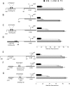
Autoregulation of virB and virF is dependent on cis-acting VirB binding sites. Summaries of the structures of various derivatives of the virB promoter region (A to D) and of the virF promoter region (E to G) are presented at the left; levels of expression of the virB-gfp (A to D) and the virF-gfp (E to G) transcriptional reporter fusions are given at the right. The data shown are relative to the level of fluorescence of a negative-control strain containing only the pZEP08 vector (Table 2). (A to D) The native virB promoter encompassing positions −310 to +30 (A), the virB promoter from positions −80 to +30 (B), the virB promoter with mutations made to the putative VirB site (C), and the virB promoter with the VirB binding site from the icsB promoter inserted at position −80 (D). (E to G) The native virF promoter encompassing positions −380 to +60 (E), the virF promoter from positions −280 to +30 (F), and the virF promoter with mutations made to the putative VirB site (G).
Fig 4
Fig 4
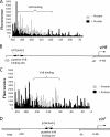
VirB requires cis-acting sequences for efficient binding at virB and at virF. (A) Plot showing the protection pattern of the virB promoter region after digestion with DNase I following incubation in the presence (black) or absence (gray) of the VirB protein. The labeled VirB site shows an area with a significant difference in the peak pattern. (B) Schematic representation of the virB promoter region shown at the same scale below the DNase I footprinting data to show the relationship of the VirB binding site to the virB transcription start site. (C) Plot showing the protection pattern of the virF promoter region after digestion with DNase I following incubation in the presence (black) or absence (gray) of the VirB protein. The labeled VirB site shows an area with a significant difference in the peak pattern. (D) Schematic representation of the virF promoter region shown at the same scale below the DNase I footprinting data to show the relationship of the VirB binding site to the virF transcription start site.
Fig 5
Fig 5
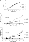
Thermal induction of the virB and virF promoters. (A) Growth of wild-type (WT) strain BS184 and its virB mutant at 30°C and 37°C over a 24-h period. (B) Expression of a virB-gfp fusion as a function of growth phase in a wild-type or virB mutant background at 30°C or 37°C. Samples were taken at regular intervals, and GFP fluorescence was measured. (C) Expression of the virF-gfp fusion as a function of growth phase in a wild-type or virB mutant background at 30°C or 37°C. Samples were taken at regular intervals, and GFP fluorescence was measured. Error bars show the standard deviations from the means.
Fig 6
Fig 6
Revised schematic overview of the interplay of transcription factors in the virulence gene regulatory cascade. The VirB protein (pentagon) binds to its own promoter, antagonizing repression mediated by H-NS (oval). The VirB protein also feeds back onto virF, binding to its promoter and antagonizing H-NS-mediated repression.

Similar articles

Cited by

References

    1. Adler B, et al. 1989. A dual transcriptional activation system for the 230-kb plasmid genes coding for virulence-associated antigens of Shigella flexneri. Mol. Microbiol. 3:627–635 - PubMed
    1. Allaoui A, Ménard R, Sansonetti PJ, Parsot C. 1993. Characterization of the Shigella flexneri ipgD and ipgF genes, which are located in the proximal part of the mxi locus. Infect. Immun. 61:1707–1714 - PMC - PubMed
    1. Allaoui A, Sansonetti PJ, Parsot C. 1993. MxiD, an outer membrane protein necessary for the secretion of the Shigella flexneri Ipa invasins. Mol. Microbiol. 7:59–68 - PubMed
    1. Andrews GP, Maurelli AT. 1992. mxiA of Shigella flexneri 2a, which facilitates export of invasion plasmid antigens, encodes a homolog of the low-calcium-response protein, LcrD, of Yersinia pestis. Infect. Immun. 60:3287–3295 - PMC - PubMed
    1. Beloin C, McKenna S, Dorman CJ. 2002. Molecular dissection of VirB, a key regulator of the virulence cascade of Shigella flexneri. J. Biol. Chem. 277:15333–15344 - PubMed

Publication types

MeSH terms

LinkOut - more resources