. 2012 Aug 31;337(6098):1091-3.
doi: 10.1126/science.1218835. Epub 2012 Jul 19.

A single progenitor population switches behavior to maintain and repair esophageal epithelium

Affiliations

A single progenitor population switches behavior to maintain and repair esophageal epithelium

David P Doupé et al. Science. .

Abstract

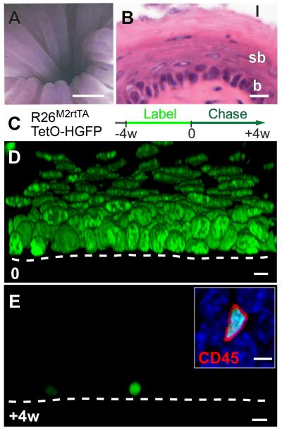
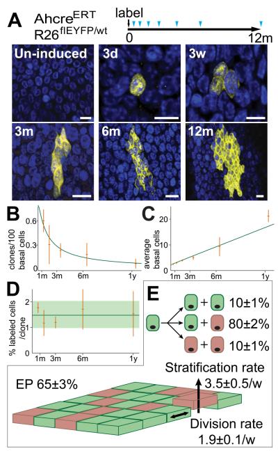
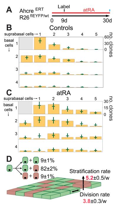
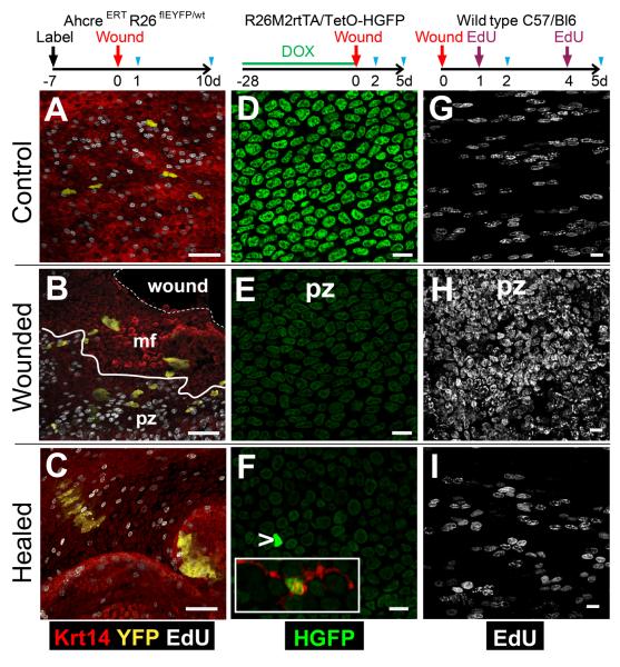
Diseases of the esophageal epithelium (EE), such as reflux esophagitis and cancer, are rising in incidence. Despite this, the cellular behaviors underlying EE homeostasis and repair remain controversial. Here, we show that in mice, EE is maintained by a single population of cells that divide stochastically to generate proliferating and differentiating daughters with equal probability. In response to challenge with all-trans retinoic acid (atRA), the balance of daughter cell fate is unaltered, but the rate of cell division increases. However, after wounding, cells reversibly switch to producing an excess of proliferating daughters until the wound has closed. Such fate-switching enables a single progenitor population to both maintain and repair tissue without the need for a "reserve" slow-cycling stem cell pool.


Figures

Fig. 1
Fig. 1. Esophageal epithelium contains no slow-cycling epithelial cells
A: Micro-endoscopy showing esophageal lumen, scale bar approx. 500μm. B: Section of epithelium, basal layer (b), suprabasal layers (sb) and lumen (l), scale bar 10μm. C: Protocol. Adult Rosa26M2rtTA/TetO-HGFP mice treated with doxycycline (DOX) express HGFP (green). Following DOX withdrawal, HGFP is diluted upon cell division, except in slow-cycling cells. D, E: Rendered confocal z stacks, showing HGFP (green) at time 0, D, and after 4w chase E (Scale bar 10μm). Dashed line indicates basement membrane. Inset shows CD45 (red) staining in HGFP retaining cell at 4w (DAPI, blue; Scale bar 5μm).
Fig. 2
Fig. 2. Proliferating cell fate in esophageal epithelium
A: Protocol: Clonal labeling was induced in AhcreERTR26flEYFP/wt mice and analyzed at intervals from 3 days to 1 year (triangles). Images are rendered confocal Z stacks of the basal layer showing typical clones at times indicated. EYFP is yellow and DAPI blue. Scale bars 10μm. B to D: Clone quantification. B, C: clone density and average clone size (basal cells). Observed values (orange) with error bars (s.e.m.), green curves show predictions of model (E). D: average % labeled basal cells at indicated time points (orange), error bars indicate s.e.m. Green line and shading show average and s.e.m. across all time points. E: Cell Fate in EE. Basal layer comprises 65% functionally equivalent esophageal progenitors (EP, green, dividing at a rate of 1.9/week, (consistent with the rate of dilution of HGFP, Fig. S1G) and 35% post-mitotic cells (pink) which stratify (arrow) at a rate of 3.5/week. 10% of EP divisions generate two EP daughters, 10% two differentiated daughters and 80% one of each fate. Values are optimal fits with 95% plausible intervals.
Fig. 3
Fig. 3. alltrans Retinoic Acid (atRA) treatment of EE
A: Protocol (see text). B and C: Size distribution of multi-cellular clones containing at least one basal cell in control (B, 307 clones), and atRA treated (C, 300 clones) EE. Green bars indicate 95% plausible fit to models in Fig. 2E (control) and 3D (atRA). D: Optimal fit during atRA treatment; proliferation and differentiation rates (red) increase compared with control.
Fig. 4
Fig. 4. Response of EP to wounding
Cartoons show protocols, blue triangles indicate sampling. A to C: Wounding of clonally labeled mice. Confocal z stacks, 1 (B) or 10 (C) days post-biopsy. mf indicates migrating front; pz, proliferative zone; solid line shows pz-mf boundary; dashed line, wound margin. A: day 1 unwounded control. EYFP is yellow, keratin 14 (Krt14) red and EdU grayscale. Scale bars 50μm. D to F: Dilution of HGFP. Confocal z stacks from unwounded control day 2 (D) and wounded mice at 2 (E) and 5 (F) days post-biopsy, showing HGFP (green). Arrow indicates HGFP bright cell (overexposed to reveal remaining cells), such cells stained for CD45 (red, inset). Scale bars 10μm. G to I: Cell proliferation. Confocal z stacks from unwounded control at 2 days (G) and experimental mice at 2 (H) and 5 (I) days post-biopsy stained for EdU (grayscale). Scale bars 10μm.

Comment in

Similar articles

Cited by

References

    1. Tumbar T, et al. Science. 2004 Jan 16;303:359. - PMC - PubMed
    1. Barker N, et al. Cell Stem Cell. 2010 Jan 8;6:25. - PubMed
    1. Barker N, et al. Nature. 2007 Oct 25;449:1003. - PubMed
    1. Jaks V, et al. Nat Genet. 2008 Nov;40:1291. - PubMed
    1. Goetsch E. Am J Anat. 1910;10:1.

Publication types

MeSH terms