. 2012 Oct;53(10):2214-2225.
doi: 10.1194/jlr.D026468. Epub 2012 Jul 20.

A novel biotinylated lipid raft reporter for electron microscopic imaging of plasma membrane microdomains

Affiliations

A novel biotinylated lipid raft reporter for electron microscopic imaging of plasma membrane microdomains

Kimberly J Krager et al. J Lipid Res. 2012 Oct.

Abstract

The submicroscopic spatial organization of cell surface receptors and plasma membrane signaling molecules is readily characterized by electron microscopy (EM) via immunogold labeling of plasma membrane sheets. Although various signaling molecules have been seen to segregate within plasma membrane microdomains, the biochemical identity of these microdomains and the factors affecting their formation are largely unknown. Lipid rafts are envisioned as submicron membrane subdomains of liquid ordered structure with differing lipid and protein constituents that define their specific varieties. To facilitate EM investigation of inner leaflet lipid rafts and the localization of membrane proteins therein, a unique genetically encoded reporter with the dually acylated raft-targeting motif of the Lck kinase was developed. This reporter, designated Lck-BAP-GFP, incorporates green fluorescent protein (GFP) and biotin acceptor peptide (BAP) modules, with the latter allowing its single-step labeling with streptavidin-gold. Lck-BAP-GFP was metabolically biotinylated in mammalian cells, distributed into low-density detergent-resistant membrane fractions, and was readily detected with avidin-based reagents. In EM images of plasma membrane sheets, the streptavidin-gold-labeled reporter was clustered in 20-50 nm microdomains, presumably representative of inner leaflet lipid rafts. The utility of the reporter was demonstrated in an investigation of the potential lipid raft localization of the epidermal growth factor receptor.


Figures

Fig. 1.
Fig. 1.
Schematic structure of a metabolically biotinylated lipid raft reporter, Lck-BAP-GFP. For the purpose of imaging lipid raft microdomains in cellular plasma membranes, a genetically encoded lipid raft-targeted reporter construct was developed that is metabolically biotinylated and thus detectible with avidin reagents. Schematic structure of the Lck-BAP-GFP reporter shows: Lck-N, the N-terminal lipid raft-targeting sequence of the Lck kinase with sites of myristoylation and dual palmitoylation indicated; BAP, the biotin acceptor peptide sequence that is metabolically biotinylated by endogenous mammalian biotin ligases; and GFP, enhanced green fluorescent protein module for detection by fluorescence microscopy.
Fig. 2.
Fig. 2.
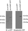
Expression and metabolic biotinylation of the Lck-BAP-GFP reporter. MDA-MB-468 breast cancer cells were lysed 24 h after transfection with the pLck-GFP or pLck-BAP-GFP vector, and cell lysate samples (15 μg protein) were subjected to SDS-PAGE and immunoblotting with anti-GFP (left). Both the 32 kDa Lck-GFP and 45 kDa Lck-BAP-GFP reporters were detected. Specific biotinylation of Lck-BAP-GFP was shown by blotting with a streptavidin-HRP conjugate and enhanced chemiluminescence detection (right).
Fig. 3.
Fig. 3.
Fluorescence imaging of the genetically encoded Lck-BAP-GFP reporter expressed in cultured cells. A: Cultured COS1 cells expressing Lck-BAP-GFP were fixed 48 h after transfection, and the GFP signal was imaged by TIRF microscopy. Shown is a representative image in which significant expression of the reporter proximal to the cell surface is evident. B: MCF7 breast cancer cells expressing Lck-BAP-GFP were imaged by laser scanning confocal microscopy. A 0.8 μm transverse optical section of a representative Lck-BAP-GFP-expressing cell shows GFP fluorescence localized primarily in the peripheral cellular membrane. Scale bars: 25 μm.
Fig. 4.
Fig. 4.
FRAP analysis of the diffusional mobility of Lck-derived reporters in the cellular plasma membrane. Cultured MCF7 cells were transfected with an Lck-GFP or Lck-BAP-GFP expression vector as indicated, and the diffusional mobility of the reporters was assessed by FRAP (see Materials and Methods). A: Representative image showing the plasma membrane fluorescence of the Lck-BAP-GFP reporter immediately after photobleaching of a 5 μm circular area. Superimposed are the bleached disk (white circle) and control (surrounding white tracing) ROIs used in the FRAP analysis. B: Quantitative analysis of the FRAP recovery for the experiment depicted in A. Integrated fluorescence intensities within the bleached and control ROIs and the corrected bleached ROI intensity data are shown. Curve fitting of the corrected data yielded the best-fit theoretical recovery curve depicted and estimates for the diffusion constant, D, and mobile fraction. C and D: Values of D (C) and the mobile fraction (D) from individual FRAP experiments are graphed in aggregate, with the mean values and SEM indicated by bars.
Fig. 5.
Fig. 5.
Identification of Lck-BAP-GFP in low-density detergent-insoluble lipid raft fractions of cellular membranes. In a conventional lipid raft fractionation protocol, MDA-MB-231 cells expressing the Lck-BAP-GFP reporter were suspended in ice-cold 1% Triton X-100, and the suspension was subjected to density gradient flotation centrifugation (see Materials and Methods). Gradient fractions were numbered in order of increasing density, and the presence of membrane markers in the fractions assessed by SDS-PAGE and immunoblotting with specific antibodies. The biotinylated Lck-BAP-GFP reporter was detected by blotting with a streptavidin-HRP conjugate (SA-HRP). The traditional lipid raft markers caveolin-1 (Cav-1, 22 kDa) and flotillin-2 (Flot-2, 47 kDa) were identified in low-density detergent-insoluble fractions, whereas the EGFR (180 kDa) and the bulk membrane (non-raft) marker transferrin receptor (TfR, 85 kDa) were found in the high-density solubilized protein fractions. Streptavidin-HRP blotting showed the presence of the biotinylated Lck-BAP-GFP marker (45 kDa) in lipid raft and solubilized fractions and the presence of previously identified endogenously biotinylated mitochondrial proteins of 75 and 125 kDa in the solubilized fractions.
Fig. 6.
Fig. 6.
EM imaging of the streptavidin-gold-labeled Lck-BAP-GFP reporter in plasma membrane sheets. Cellular plasma membrane sheets from MDA-MB-468 breast cancer cells expressing either the Lck-BAP-GFP or Lck-GFP reporter were isolated and prepared as for standard immunogold labeling but labeled with a 6 nm streptavidin-gold conjugate. Labeled membranes were imaged by transmission EM at 12,000× (see Materials and Methods). A: EM image of a streptavidin-gold-labeled plasma membrane sheet from an Lck-BAP-GFP-expressing cell. Submicron-sized clusters of the gold-labeled reporter were evident (see arrow). B: EM image of streptavidin-gold-labeled membrane of a control cell expressing the Lck-GFP reporter. C: Quantification of labeling of Lck-BAP-GFP-expressing versus Lck-GFP-expressing (control) cells in repeated experiments. Shown are the mean and SEM for 11 experiments in which particles in equivalent membrane areas were counted. D: Representative Ripley's K function analysis of the clustering of gold particles in EM images of labeled Lck-BAP-GFP-expressing MDA-MB-468 cell membranes. The image analyzed (a cropped version of which is shown in A) corresponded to a total membrane area of 1443 nm × 1443 nm. The graph displays the evaluated function SQRT[K(r)/π] – r for the particle distribution (solid line). Also shown are the envelopes of the maximum and minimum deviations of the evaluated functions (dotted lines) for 99 randomly generated distributions of the same number of particles. A nonrandom distribution was evident with clustering on the dimension of 20–50 nm.
Fig. 7.
Fig. 7.
Double-labeling of the Lck-BAP-GFP reporter with streptavidin-gold and conventional immunogold reagents. Membrane sheets from MDA-MB-468 cells expressing Lck-BAP-GFP were labeled with a GFP-specific rabbit polyclonal antibody/10 nm gold-conjugated secondary antibody pair and a 6 nm gold-conjugated streptavidin reagent. A: Representative EM image (cropped version of the larger analyzed image) of 6 nm streptavidin-gold (arrow) and anti-GFP/10 nm gold (arrowhead) labeling in a plasma membrane sheet (12,000×). B–D: Ripley's K function plots (solid lines) characterizing the spatial distributions of streptavidin-gold (B) and anti-GFP/immunogold (C) labeling and a bivariate K function plot (solid line) characterizing the co-clustering of streptavidin-gold and anti-GFP/immunogold (D). The envelopes of the evaluated K functions for 99 randomly generated distributions of the same numbers of particles are also indicated (dotted lines). Both streptavidin-gold and anti-GFP/immunogold individually showed clustering on the scale of 20–50 nm, and the bivariate analysis showed a co-clustering of the two labels on a similar size scale.
Fig. 8.
Fig. 8.
Application of the Lck-BAP-GFP reporter in assessing the potential lipid raft localization of the EGFR. Membrane sheets from MDA-MB-468 cells expressing a high level of endogenous EGFR and the ectopic Lck-BAP-GFP lipid raft marker were analyzed by EM after gold labeling of the EGFR and Lck-BAP-GFP proteins. The EGFR was immunolabeled with a rabbit monoclonal antibody and 10 nm gold-conjugated secondary antibody, and the biotinylated raft marker was labeled with a 6 nm gold-conjugated streptavidin reagent. A: Representative transmission EM image (cropped version of the larger analyzed image) of 10 nm gold-labeled EGFR (arrowhead) and 6 nm gold-labeled Lck-BAP-GFP (arrow) proteins in a plasma membrane sheet (12,000×). B and C: Ripley's K function analysis of the clustering of the labeled Lck-BAP-GFP reporter (B) and EGFR (C). D: The evaluated bivariate K function (solid line) for the Lck-BAP-GFP and EGFR particle distributions in the representative image fell largely within the envelope of 99 simulated random distributions (dotted lines), which indicated little tendency for the EGFR and raft reporter to co-cluster.

Similar articles

Cited by

References

    1. Simons K., Toomre D. 2000. Lipid rafts and signal transduction. Nat. Rev. Mol. Cell Biol. 1: 31–39 - PubMed
    1. Tian T., Harding A., Inder K., Plowman S., Parton R. G., Hancock J. F. 2007. Plasma membrane nanoswitches generate high-fidelity Ras signal transduction. Nat. Cell Biol. 9: 905–914 - PubMed
    1. Wilson B. S., Pfeiffer J. R., Oliver J. M. 2002. FcepsilonRI signaling observed from the inside of the mast cell membrane. Mol. Immunol. 38: 1259–1268 - PubMed
    1. Wilson B. S., Steinberg S. L., Liederman K., Pfeiffer J. R., Surviladze Z., Zhang J., Samelson L. E., Yang L. H., Kotula P. G., Oliver J. M. 2004. Markers for detergent-resistant lipid rafts occupy distinct and dynamic domains in native membranes. Mol. Biol. Cell. 15: 2580–2592 - PMC - PubMed
    1. Nicolau D. V., Jr, Burrage K., Parton R. G., Hancock J. F. 2006. Identifying optimal lipid raft characteristics required to promote nanoscale protein-protein interactions on the plasma membrane. Mol. Cell. Biol. 26: 313–323 - PMC - PubMed

Publication types

Substances