. 2012 Nov;135(Pt 11):3251-64.
doi: 10.1093/brain/aws178. Epub 2012 Jul 20.

The tissue-type plasminogen activator-plasminogen activator inhibitor 1 complex promotes neurovascular injury in brain trauma: evidence from mice and humans

Affiliations

The tissue-type plasminogen activator-plasminogen activator inhibitor 1 complex promotes neurovascular injury in brain trauma: evidence from mice and humans

Maithili Sashindranath et al. Brain. 2012 Nov.

Abstract

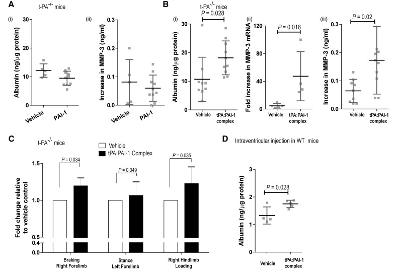
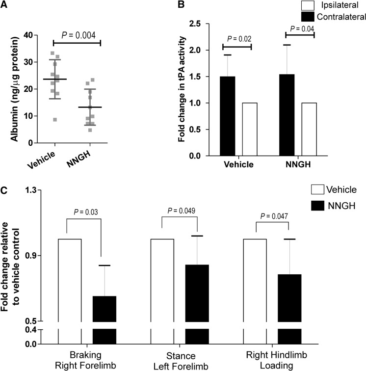
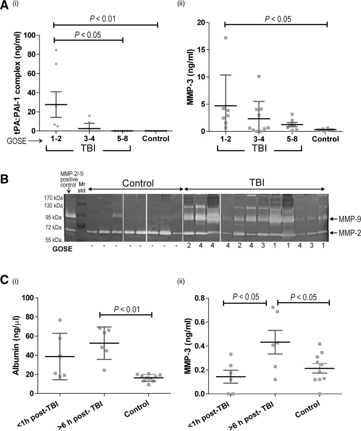
The neurovascular unit provides a dynamic interface between the circulation and central nervous system. Disruption of neurovascular integrity occurs in numerous brain pathologies including neurotrauma and ischaemic stroke. Tissue plasminogen activator is a serine protease that converts plasminogen to plasmin, a protease that dissolves blood clots. Besides its role in fibrinolysis, tissue plasminogen activator is abundantly expressed in the brain where it mediates extracellular proteolysis. However, proteolytically active tissue plasminogen activator also promotes neurovascular disruption after ischaemic stroke; the molecular mechanisms of this process are still unclear. Tissue plasminogen activator is naturally inhibited by serine protease inhibitors (serpins): plasminogen activator inhibitor-1, neuroserpin or protease nexin-1 that results in the formation of serpin:protease complexes. Proteases and serpin:protease complexes are cleared through high-affinity binding to low-density lipoprotein receptors, but their binding to these receptors can also transmit extracellular signals across the plasma membrane. The matrix metalloproteinases are the second major proteolytic system in the mammalian brain, and like tissue plasminogen activators are pivotal to neurological function but can also degrade structures of the neurovascular unit after injury. Herein, we show that tissue plasminogen activator potentiates neurovascular damage in a dose-dependent manner in a mouse model of neurotrauma. Surprisingly, inhibition of activity following administration of plasminogen activator inhibitor-1 significantly increased cerebrovascular permeability. This led to our finding that formation of complexes between tissue plasminogen activator and plasminogen activator inhibitor-1 in the brain parenchyma facilitates post-traumatic cerebrovascular damage. We demonstrate that following trauma, the complex binds to low-density lipoprotein receptors, triggering the induction of matrix metalloproteinase-3. Accordingly, pharmacological inhibition of matrix metalloproteinase-3 attenuates neurovascular permeability and improves neurological function in injured mice. Our results are clinically relevant, because concentrations of tissue plasminogen activator: plasminogen activator inhibitor-1 complex and matrix metalloproteinase-3 are significantly elevated in cerebrospinal fluid of trauma patients and correlate with neurological outcome. In a separate study, we found that matrix metalloproteinase-3 and albumin, a marker of cerebrovascular damage, were significantly increased in brain tissue of patients with neurotrauma. Perturbation of neurovascular homeostasis causing oedema, inflammation and cell death is an important cause of acute and long-term neurological dysfunction after trauma. A role for the tissue plasminogen activator-matrix metalloproteinase axis in promoting neurovascular disruption after neurotrauma has not been described thus far. Targeting tissue plasminogen activator: plasminogen activator inhibitor-1 complex signalling or downstream matrix metalloproteinase-3 induction may provide viable therapeutic strategies to reduce cerebrovascular permeability after neurotrauma.


Figures

Figure 1
Figure 1
t-PA activity is associated with increased neurovascular permeability, lesion volume and gelatinolytic activity after TBI. (A) Evan’s blue extravasation is increased in T4 transgenic relative to T4 wild-type mice at 3 h post-TBI. t-PA−/− mice are protected from cerebrovascular damage relative to wild-type mice (Wt); T4 transgenic mice (injured n = 5, sham n = 4); T4 wild-type mice (injured n = 8, sham n = 3); wild-type mice (injured n = 6, sham n = 4); t-PA−/− mice (injured n = 7, sham n = 8). (B) T4 mice have larger lesion volumes relative to T4 wild-type mice 24 h after severe trauma; T4 transgenic and T4 wild-type injured (n = 8), sham (n = 6). (C) T4 mice have increased Rotarod deficit relative to T4 wild-type at 24 h post-severe TBI; injured T4 transgenic and T4 wild-type mice (n = 8), sham T4 mice (n = 4), T4 wild-type mice (n = 3). (D) Gelatinolytic activity is increased in T4 mice relative to T4 wild-type mice at 3 h post-TBI. (i) A t-PA-dependent increase in pro-MMP9 in the ipsilateral (ipsi) relative to the contralateral cortex (contra) in injured T4 and T4 wild-type mice; MMP-2 activity is increased equally in both T4 and T4 wild-type ipsilateral cortex; MMP2/9 positive control is conditioned media from HT1080 fibrosarcoma cells. (ii) Fold change in pro-MMP9 activity i.e. increase in activity in injured normalized to the baseline activity value of sham mice; T4 transgenic and T4 wild-type mice (injured n = 6); sham T4 (n = 4), T4 wild-type (n = 3). (E) Temporal profile of gelatinolytic activity shows a selective increase at 3 h following TBI and return to baseline levels by 24 h post-TBI in the ipsilateral relative to the contralateral cortex as determined by extent of cleavage of fluorescein isothiocyanate–gelatin in wild-type mice; n = 5. Data expressed as mean ± SD. T4-Wt = T4 wild-type.
Figure 2
Figure 2
Injection of PAI1 exacerbates neurovascular damage via LDLRs. (A) Intracortical injection of stable mouse PAI1 post-TBI in wild-type mice (i) inhibits t-PA activity within the ipsilateral cortex as measured by an S2251-based amidolytic assay (Sashindranath et al., 2011); receptor associated protein does not interfere with the inhibitory action of PAI1 against t-PA; n = 8–10 (data depicted as fold-change in t-PA activity the ipsilateral cortex normalized to the t-PA activity in the contralateral cortex) and (ii) causes a significant increase in albumin extravasation in the ipsilateral relative to the contralateral cortex at 3 h post-TBI; this increase is blocked when receptor associated protein is co-injected with PAI1. [A(iii)] Concentration of MMP3 antigen in the ipsilateral cortex increases when PAI1 is injected, and decreases when receptor associated protein is co-injected with PAI1; n = 9 PAI1, n = 8 PAI1 + receptor associated protein, n = 10 vehicle. [B(i)]: Intra-cortical injection of the stable human PAI1 R76E mutant after TBI in wild-type mice inhibits t-PA activity (ii) PAI1 R76E mutant has no effect on albumin extravasation or (iii) MMP3 concentrations at 3 h post-TBI when injected intracortically; n = 7; PAI1 R76E, n = 8 vehicle. Data expressed as mean ± SD. RAP = receptor associated protein.
Figure 3
Figure 3
t-PA:PAI1 complex formation promotes neurovascular damage after TBI. (A) Intracortical injection of stable PAI1 in t-PA−/− mice post-TBI does not alter (i) albumin extravasation or (ii) MMP3 antigen levels relative to vehicle controls at 3 h post-TBI (PAI1 n = 5, vehicle n = 10). [B(i)] Intracortical injection of the preformed t-PA:PAI1 complex into the ipsilateral cortex of t-PA−/− mice increases vascular permeability (t-PA:PAI1 complex n = 9; vehicle n = 10); (ii) MMP3 messenger RNA (t-PA:PAI1 complex n = 5, vehicle n = 4); and (iii) MMP3 antigen levels 3 h post-trauma (t-PA:PAI1 complex n = 9; vehicle n = 10). (C) DigiGait™ analysis shows a significant increase in post:pre-injury ratio of deceleration in the right forelimb, stance in the left forelimb as well as in right hind-limb loading, indicating a greater behavioural deficit following intracortical injection of the t-PA:PAI1 complex after TBI; normalized to the post:pre-injury ratio of these parameters in vehicle injected t-PA−/− animals; n = 11. (D) Intraventricular injection of the t-PA:PAI1 complex (500 nM in 1.5 µl) in wild-type mice significantly increases albumin extravasation at 1 h post-injection (t-PA:PAI1 complex n = 5, vehicle n = 4). Data expressed as mean ± SD. WT = wild-type.
Figure 4
Figure 4
MMP3 mediates neurovascular breakdown at 3 h post-TBI. (A) Oral administration of the MMP3 inhibitor NNGH into wild-type mice reduces albumin extravasation at 3 h post-TBI. (B) NNGH administration does not alter the TBI-induced increase in t-PA activity in the ipsilateral cortex. (C) NNGH causes a significant improvement in post : pre-injury ratio of braking in the right forelimb, stance in the left forelimb as well as in right hindlimb loading, indicating improved neurological outcome; data normalized to the post : pre-injury ratio of these parameters in vehicle-gavaged control mice, n = 10. Data expressed as mean ± SD.
Figure 5
Figure 5
t-PA:PAI1 complex and MMP3 are elevated after injury in CSF of trauma patients, and concentrations of MMP3 and albumin are increased in the injured cortex of human TBI cases. [A(i)] t-PA:PAI1 complex concentration is elevated in patients with GOS-E score 1–2 (death, vegetative state; n = 7), low in patients with GOS-E score 3–4 (severe disability; n = 10) and undetectable in patients with GOS-E score of 5–8 (moderate disability to good recovery; n = 6) and in all control patients (n = 8); t-PA:PAI1 complex levels correlate with neurological outcome (r = −0.539; P = 0.0079, see Supplementary Fig. 1A). [A(ii)] MMP3 concentration in CSF also increases with injury severity, and correlates with the t-PA:PAI1 complex (r = 0.585; P = 0.0006, Supplementary Fig. 1B). (B) Gelatin zymogram shows marked elevation in MMP9 and likely MMP9:TIMP-1 complexes but not MMP2 activity in CSF of 12 patients with TBI (GOS-E score 1–4; indicated below gel). MMP9 is absent or weakly detected in eight non-TBI control patients. MMP2/9 positive control is conditioned media of HT-1080 fibrosarcoma cells. The results presented comprise images from four separate (non-contiguous) zymograms of CSF samples from control and patients with TBI; control and TBI sample lanes have been grouped for clarity. The individual gels are separated by a white line. [C(i)] Increased albumin content in brain tissue of patients with TBI compared with non-TBI control cases (n = 10) within 1 h of injury (n = 6) and a further increased in patients who survived ≥6 h post-injury (n = 6) (Table 1). (ii) MMP3 antigen levels in the same cortical tissue are significantly elevated at ≥6 h post-injury compared with non-TBI control patients. Data expressed as mean ± SD.

References

    1. Alvarez JI, Cayrol R, Prat A. Disruption of central nervous system barriers in multiple sclerosis. Biochim Biophys Acta. 2011;1812:252–64. - PubMed
    1. Armstead WM, Kiessling JW, Riley J, Cines DB, Higazi AA. tPA contributes to impaired NMDA cerebrovasodilation after traumatic brain injury through activation of JNK MAPK. Neurol Res. 2011;33:726–33. - PMC - PubMed
    1. Armstead WM, Nassar T, Akkawi S, Smith DH, Chen XH, Cines DB, et al. Neutralizing the neurotoxic effects of exogenous and endogenous tPA. Nat Neurosci. 2006;9:1150–5. - PubMed
    1. Candelario-Jalil E, Yang Y, Rosenberg GA. Diverse roles of matrix metalloproteinases and tissue inhibitors of metalloproteinases in neuroinflammation and cerebral ischemia. Neuroscience. 2009;158:983–94. - PMC - PubMed
    1. Dietzmann K, von Bossanyi P, Krause D, Wittig H, Mawrin C, Kirches E. Expression of the plasminogen activator system and the inhibitors PAI-1 and PAI-2 in posttraumatic lesions of the CNS and brain injuries following dramatic circulatory arrests: an immunohistochemical study. Pathol Res Prac. 2000;196:15–21. - PubMed

Publication types

MeSH terms

Substances