. 2012 Aug;2(8):722-35.
doi: 10.1158/2159-8290.CD-12-0014. Epub 2012 Jul 19.

IDO is a nodal pathogenic driver of lung cancer and metastasis development

Affiliations

IDO is a nodal pathogenic driver of lung cancer and metastasis development

Courtney Smith et al. Cancer Discov. 2012 Aug.

Abstract

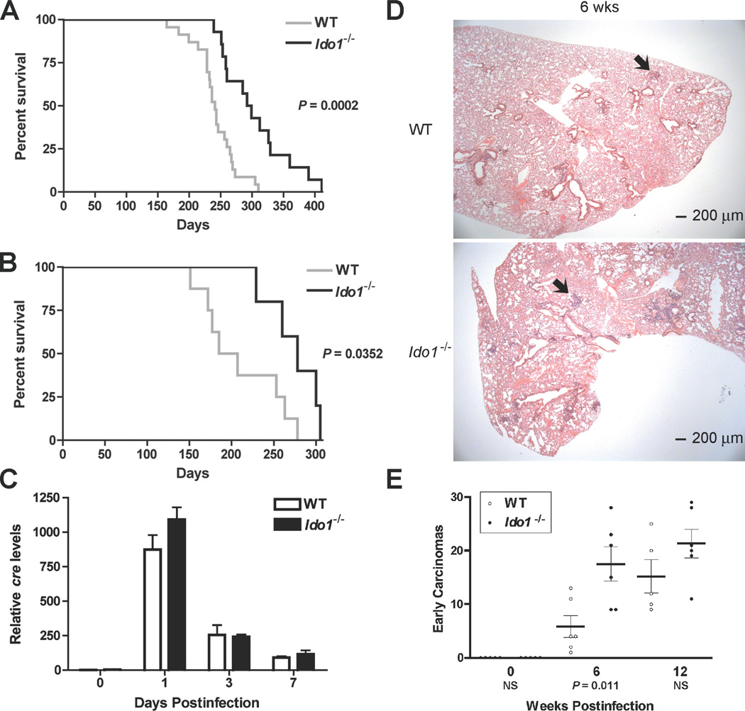
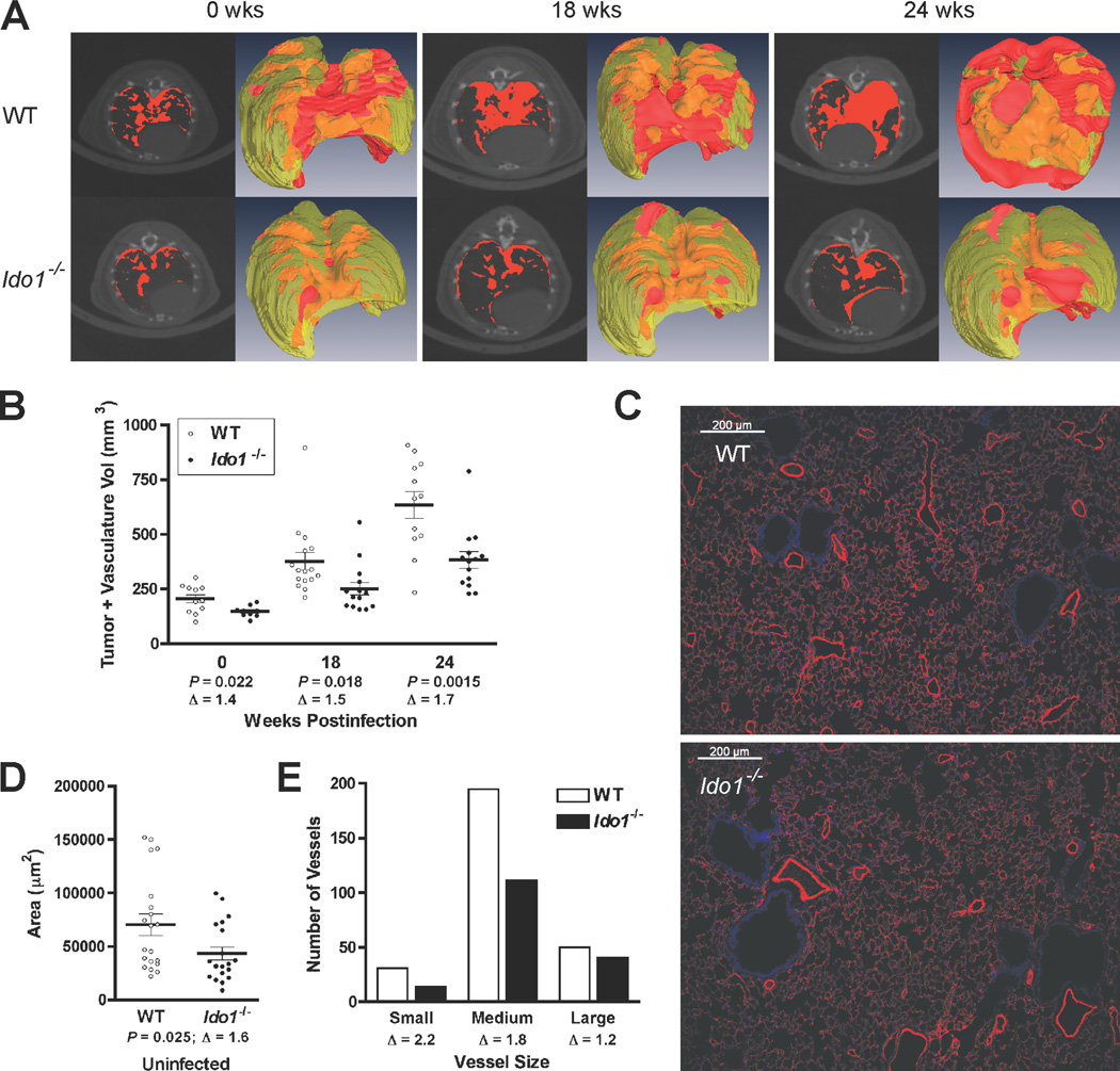
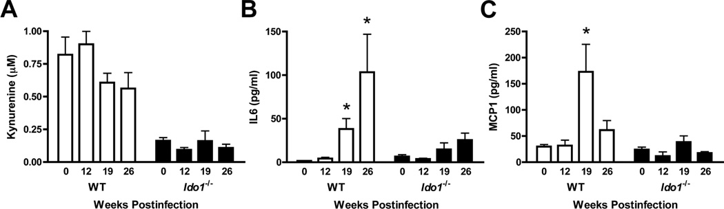
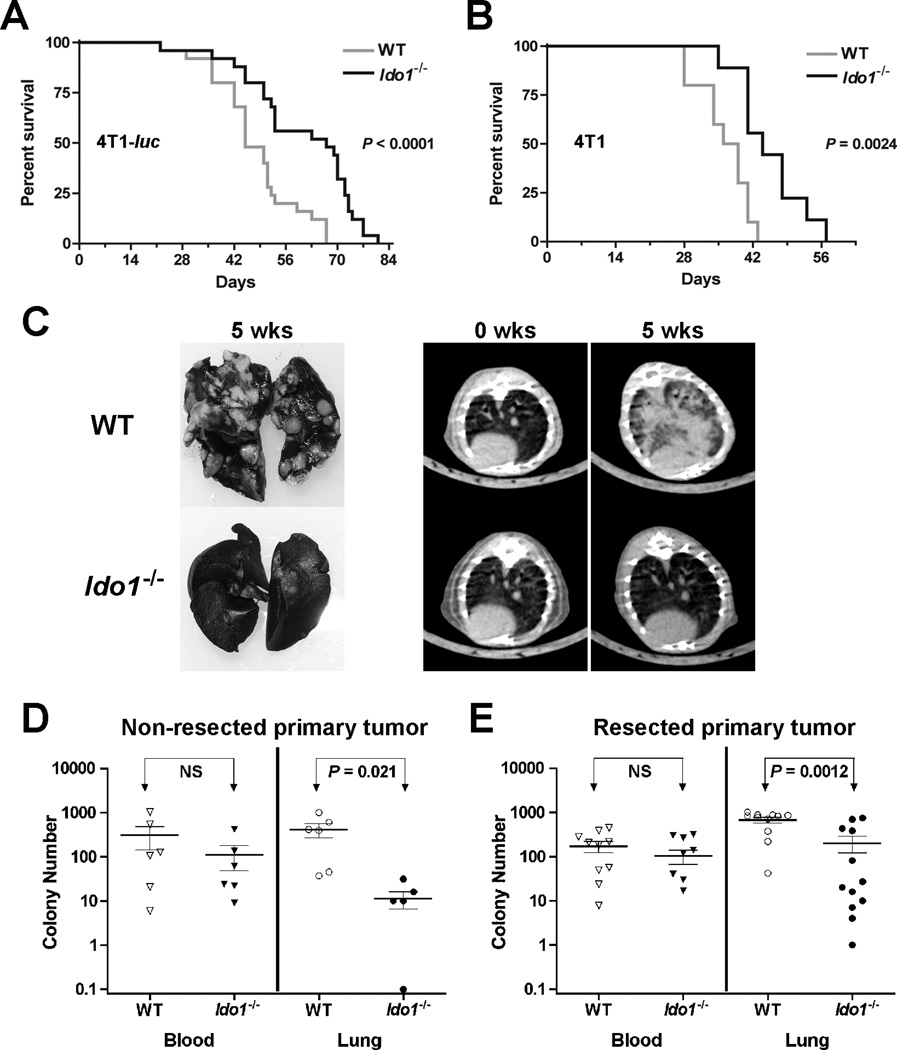
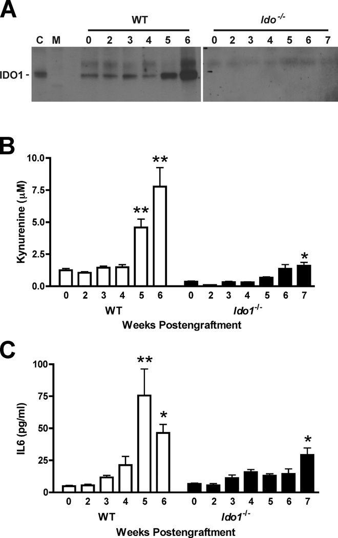
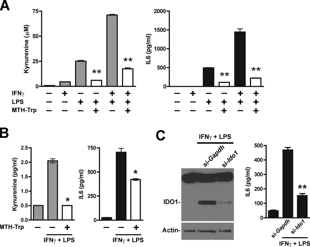
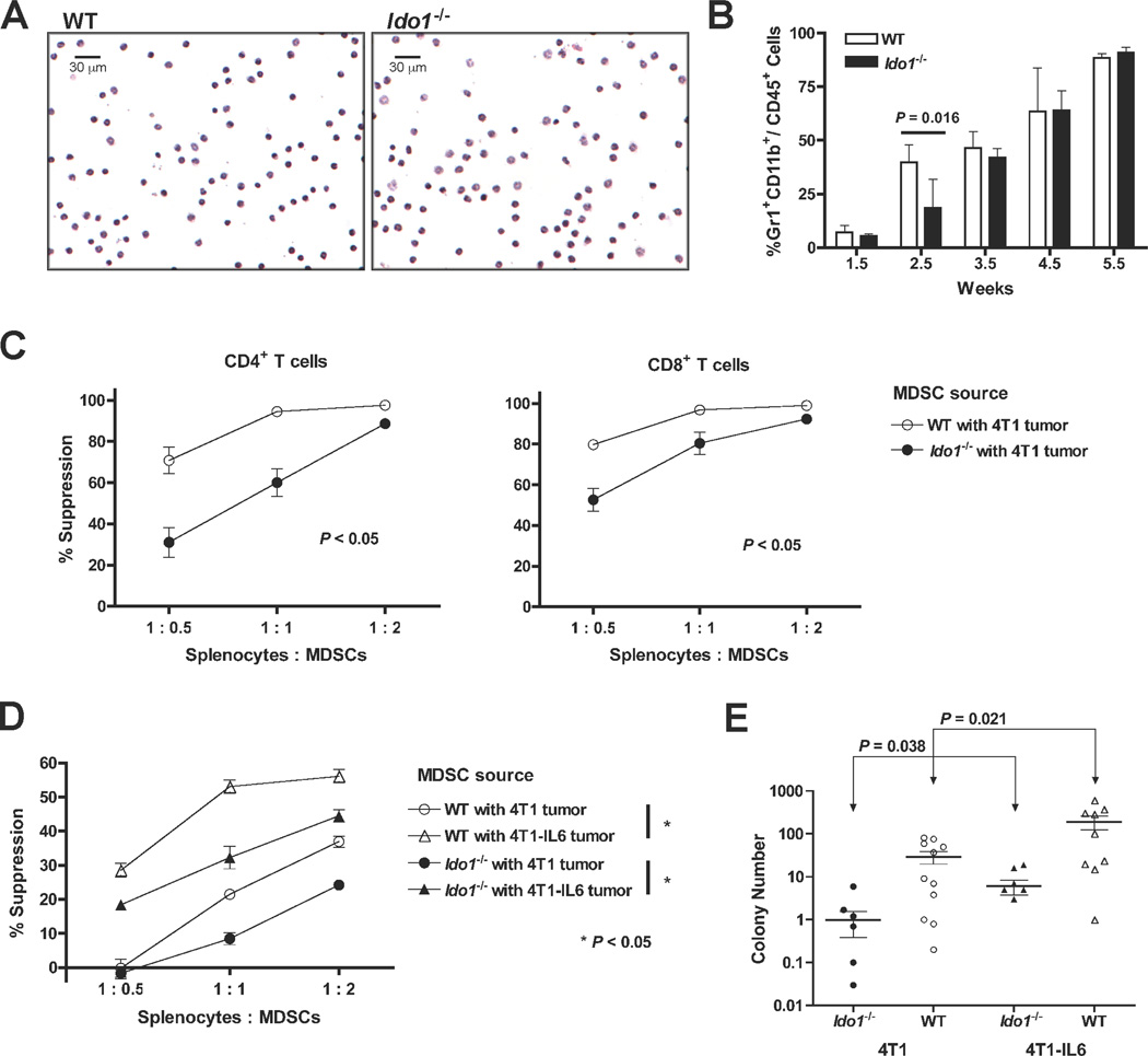
Indoleamine 2,3-dioxygenase (IDO) enzyme inhibitors have entered clinical trials for cancer treatment based on preclinical studies, indicating that they can defeat immune escape and broadly enhance other therapeutic modalities. However, clear genetic evidence of the impact of IDO on tumorigenesis in physiologic models of primary or metastatic disease is lacking. Investigating the impact of Ido1 gene disruption in mouse models of oncogenic KRAS-induced lung carcinoma and breast carcinoma-derived pulmonary metastasis, we have found that IDO deficiency resulted in reduced lung tumor burden and improved survival in both models. Micro-computed tomographic (CT) imaging further revealed that the density of the underlying pulmonary blood vessels was significantly reduced in Ido1-nullizygous mice. During lung tumor and metastasis outgrowth, interleukin (IL)-6 induction was greatly attenuated in conjunction with the loss of IDO. Biologically, this resulted in a consequential impairment of protumorigenic myeloid-derived suppressor cells (MDSC), as restoration of IL-6 recovered both MDSC suppressor function and metastasis susceptibility in Ido1-nullizygous mice. Together, our findings define IDO as a prototypical integrative modifier that bridges inflammation, vascularization, and immune escape to license primary and metastatic tumor outgrowth.

Significance: This study provides preclinical, genetic proof-of-concept that the immunoregulatory enzyme IDO contributes to autochthonous carcinoma progression and to the creation of a metastatic niche. IDO deficiency in vivo negatively impacted both vascularization and IL-6–dependent, MDSC-driven immune escape, establishing IDO as an overarching factor directing the establishment of a protumorigenic environment.


Figures

Figure 1
Figure 1
IDO-deficiency extends the survival of mice with KRAS-induced lung adenocarcinomas despite an elevated number of early lesions. (A) Kaplan-Meier survival curves for cohorts of Lox-KrasG12D (N = 23) and Ido1−/− Lox-KrasG12D (N = 14) mice infected with 2.5 × 10⁷ PFU Ad-cre virus. (B) Kaplan-Meier survival curves for cohorts of Lox-KrasG12D (N = 8) and Ido1−/− Lox-KrasG12D (N = 5) mice infected with 1.25 × 10⁸ PFU Ad-cre virus. Significance for both data sets was assessed by 2-group log-rank test at P < 0.05. (C) Total lung DNA prepared from 3 mice per time point was analyzed for the presence of the viral cre gene by real time PCR at 0, 1, 3, 7 days postinfection. Relative cre levels determined from this analysis are plotted as means ± SEM. (D) Representative H&E stained sections depicting the observed difference in early lesions between the lungs of Lox-KrasG12D and I d o 1−/− Lox-KrasG12D mice at 6 weeks postinfection. (E) Quantitative histopathological assessment of lesion frequency in H&E stained sections of lung biopsies from Lox-KrasG12D and Ido1−/− Lox-KrasG12D mice at 6wk and 12wk postinfection (N ≥ 5). The number of lesions identifiable under low magnification within a defined region of each specimen are graphed on the scatter plot with the means ± SEM. Significance was determined by two-tailed Student’s t test at P < 0.05) (NS; not significant).
Figure 2
Figure 2
IDO-deficiency impairs the outgrowth of overt lung adenocarcinomas and reduces normal pulmonary vascularization. (A) Representative axial micro-CT images and 3-D reconstructions of Lox-KrasG12D and Ido1−/− Lox-KrasG12D mouse lungs acquired at 0, 18 and 24 weeks post-infection. Tumor and vasculature, which have indistinguishable X-ray densities, are designated in red in the individual CT images (left panels) or red/orange for exterior/interior location in the 3-D reconstructions (right panels). (B) Volumetric image analysis of tumor and vasculature performed on the 3-D reconstructions of lung micro-CT images. The data are graphed as a scatter plot with the means ± SEM (Δ; fold difference). Significance was determined by two-tailed Student’s t test at P < 0.05. (C) Immunofluorescent staining of blood vessels with antibody to caveolin 1 (red) and DAPI staining of nuclei (blue) in representative lung tissue specimens from WT and Ido1−/− mice. (D) Quantitation of blood vessel areas. The vessel area totals measured within each field are graphed as a scatter plot with the means ± SEM (Δ; fold difference). Significance was determined by two-tailed Student’s t test at P < 0.05. (E) Distribution of pulmonary vessels within specified size ranges. The total number of small (<500 µm2), medium (500–5000 µm2) and large (>5000 µm2) vessels identified within the defined fields evaluated in D are plotted on a bar graph (Δ; fold difference). Also see Supplementary Fig. S1C for a graph of individual vessel measurements rank ordered across the entire size range.
Figure 3
Figure 3
IDO-deficiency is associated with attenuated induction of IL6 and MCP1. (A) Kynurenine levels in the lungs of Lox-KrasG12D and Ido1−/− Lox-KrasG12D mice at 0, 12, 19, and 26 weeks post-infection (N ≥ 3) assessed by liquid chromatography-tandem mass spectroscopy analysis and plotted as the means ± SEM. (B,C) IL6 and MCP1 levels in the lungs of Lox-KrasG12D and Ido1−/− Lox-KrasG12D mice at 0, 12, 19, and 26 weeks postinfection (N ≥ 3) assessed by multiplexed cytokine bead immunoassay-based analysis and plotted as the means ± SEM with significance relative to baseline determined by 1-way ANOVA with Dunn’s test (*; P < 0.05).
Figure 4
Figure 4
IDO-deficiency delays the development of pulmonary metastases. Kaplan-Meier survival curves for cohorts of WT and Ido1−/− mice following orthotopic engraftment of 1×10⁴ (A) 4T1-luc (N = 25) or (B) 4T1 (N ≥ 9) tumor cells. Significance was assessed by 2-group log-rank test at P < 0.05. The survival benefit observed in Ido1−/− mice was independently replicated at UMBC. (C) Staining of lungs with India ink and axial images from micro-CT scans depicting the difference in pulmonary metastasis burden between WT and Ido1−/− mice at 5 weeks following orthotopic 4T1 tumor cell engraftment. At 5 weeks following (D) orthotopic engraftment of 4T1 cells (N = 6) or (E) orthotopic engraftment of 4T1 cells and resection of the primary tumor at 18 days post-engraftment (N ≥ 11), colony forming assays were performed to assess the relative tumor cell burden in the blood (neat) and lungs (1:1000). Individual data points are graphed on a log scale scatter plot with the means ± SEM and significance assessed by two-tailed Student’s t test at P < 0.05 (NS; not significant).
Figure 5
Figure 5
IDO-deficiency is associated with attenuated induction of IL6 during 4T1 tumor metastasis. (A) Evaluation of IDO1 protein levels by IP-Western blot analysis of lung tissue lysates from WT and Ido1−/− mice following orthotopic engraftment of 4T1 tumor cells at the time points in weeks indicated above each lane, [C; epididymis lysate positive control lane, M; molecular weight marker lane]. (B) Evaluation of kynurenine levels by liquid chromatography-tandem mass spectroscopy-based analysis of homogenized lung samples from WT and Ido1−/− mice following orthotopic engraftment of 4T1 tumor cells at the time points in weeks indicted for each lane. Means ± SEM (N ≥ 6) are graphed with significance relative to baseline determined by 1-way ANOVA with Dunn’s test (*; P < 0.05, **; P < 0.01). (C) IL6 level determinations from cytokine bead array immunoassay-based analysis of homogenized lung samples from WT and Ido1−/− mice following orthotopic engraftment of 4T1 tumor cells at the time points in weeks indicted for each lane. Means ± SEM (N ≥ 3) are graphed with significance relative to baseline determined by 1-way ANOVA with Dunn’s test (*; P < 0.05, **; P < 0.01).
Figure 6
Figure 6
IDO-dependent potentiation of IL6 production. (A) Supernatant from U937 cells stimulated for 24 hours with IFNγ (100 ng/ml) and/or LPS (100 ng/ml) was analyzed for kynurenine and IL6. Results from triplicate wells are plotted as the means ± SEM. Methyl thiohydantoin tryptophan (MTH-Trp, 100 µmol/l) was included during induction where indicated and significance relative to the corresponding induced level without MTH-Trp was determined by two-tailed Student’s t test (**; P ≤ 0.0001). (B) Supernatant from HL-60 cells stimulated for 24 hours with IFNγ (100 ng/ml) and LPS (100 ng/ml) was analyzed for kynurenine and IL6. Results from duplicate wells are plotted as the means ± SEM. Methyl thiohydantoin tryptophan (MTH-Trp, 100 µmol/l) was included during induction where indicated and significance relative to the corresponding induced level without MTH-Trp was determined by two-tailed Student’s t test (*; P < 0.05). (C) HL-60 cells treated in triplicate with Ido1-targeting (si-Ido1) or non-targeting (si-Gapdh) siRNAs were stimulated for 24 hours with IFNγ (100 ng/ml) and LPS (100 ng/ml). Pooled cell lysates were analyzed by Western blot analysis for IDO1 and β-actin (left panel). IDO1 induction was suppressed by approximately 89.7% as assessed by densitometry analysis and normalization to actin. Individual cell supernatants were analyzed for IL6 (right panel). The IL6 data are plotted as the means ± SEM with the significance of the difference between specific Ido1-targeting versus non-targeting results determined by two-tailed Student’s t test (**; P < 0.0001).
Figure 7
Figure 7
Attenuated MDSC suppressive activity and metastasis development in IDO-deficient mice is rescued by IL6. (A) Comparative microscopic images of H&E-stained MDSCs harvested from the blood of WT and Ido1−/− mice with primary 4T1 tumors that were not significantly different in size (12.2 ± 1.36 and 11. 5± 0.4 mm in diameter, respectively). (B) Single cell suspensions of whole lung tissues were prepared at the indicated time points following 4T1 engraftment into WT and Ido1−/− mice and evaluated by flow cytometry for MDSC infiltration by gating on CD45+ cells and analyzing the Gr1+CD11b+ cell population. Means ± SEM are graphed with significance assessed by two-tailed Student’s t test at P < 0.05. (C) Splenocytes from CD4+ TS1 (left) or CD8+ Clone 4 (right) mice were co-cultured in triplicate with cognate peptide and increasing proportions of 4T1-induced, peripheral blood MDSCs from WT or Ido1−/− mice. T cell activation was quantified by uptake of 3H-thymidine and graphed as percent suppression relative to activation in the absence of MDSCs. Significance was assessed by Wilcoxon Rank test at P < 0.05. Outcomes are representative of a minimum of 3 independent experiments. (D) Splenocytes from CD8+ Clone 4 transgenic mice were co-cultured with cognate peptide and increasing proportions of 4T1 or 4T1-IL6 tumor-induced MDSCs from WT or Ido1−/− mice for analysis as in B. Outcomes are representative of 6 independent experiments using TS1, Clone 4, or DO11.10 transgenic T cells. (E) Colony forming assays to assess the relative tumor cell burden in the lungs performed 6 weeks following intravenous injection of 4T1 or 4T1-IL6 cells into WT and Ido1−/− mice. Results are presented on log scale scatter plot with means ± SEM. Significance was assessed by two-tailed Student’s t test at P < 0.05.

Comment in

References

    1. Peek RM, Jr, Mohla S, DuBois RN. Inflammation in the genesis and perpetuation of cancer: summary and recommendations from a national cancer institute-sponsored meeting. Cancer Res. 2005;65:8583–8586. - PubMed
    1. Muller AJ, Mandik-Nayak L, Prendergast GC. Beyond immunosuppression: reconsidering indoleamine 2,3-dioxygenase as a pathogenic element of chronic inflammation. Immunotherapy. 2010;2:293–297. - PMC - PubMed
    1. Muller AJ, Scherle PA. Targeting the mechanisms of tumoral immune tolerance with small-molecule inhibitors. Nat Rev Cancer. 2006;6:613–625. - PubMed
    1. Banerjee T, DuHadaway JB, Gaspari P, Sutanto-Ward E, Munn DH, Mellor AL, et al. A key in vivo antitumor mechanism of action of natural product-based brassinins is inhibition of indoleamine 2,3-dioxygenase. Oncogene. 2008;27:2851–2857. - PubMed
    1. Hou DY, Muller AJ, Sharma MD, DuHadaway J, Banerjee T, Johnson M, et al. Inhibition of indoleamine 2,3-dioxygenase in dendritic cells by stereoisomers of 1-methyl-tryptophan correlates with antitumor responses. Cancer Res. 2007;67:792–801. - PubMed

Publication types

MeSH terms

Substances