. 2012 Sep 7;287(37):30967-74.
doi: 10.1074/jbc.M112.382507. Epub 2012 Jul 20.

Nuclear calcium signaling controls methyl-CpG-binding protein 2 (MeCP2) phosphorylation on serine 421 following synaptic activity

Affiliations

Nuclear calcium signaling controls methyl-CpG-binding protein 2 (MeCP2) phosphorylation on serine 421 following synaptic activity

Bettina Buchthal et al. J Biol Chem. .

Abstract

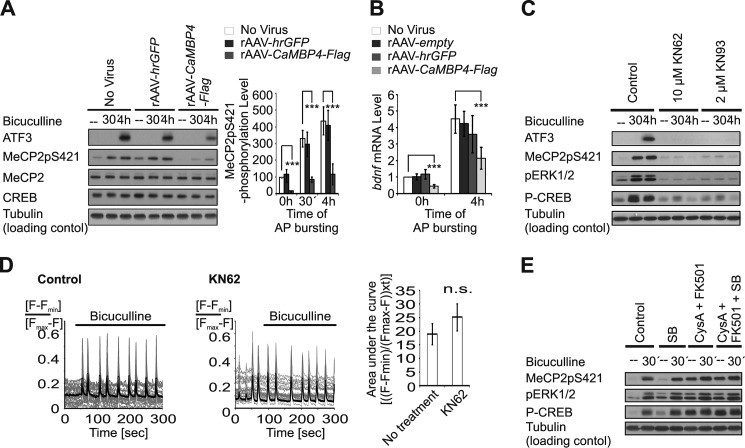
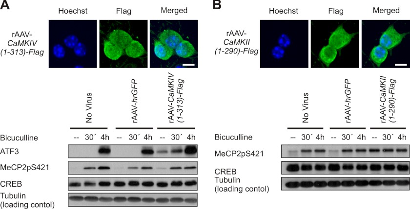
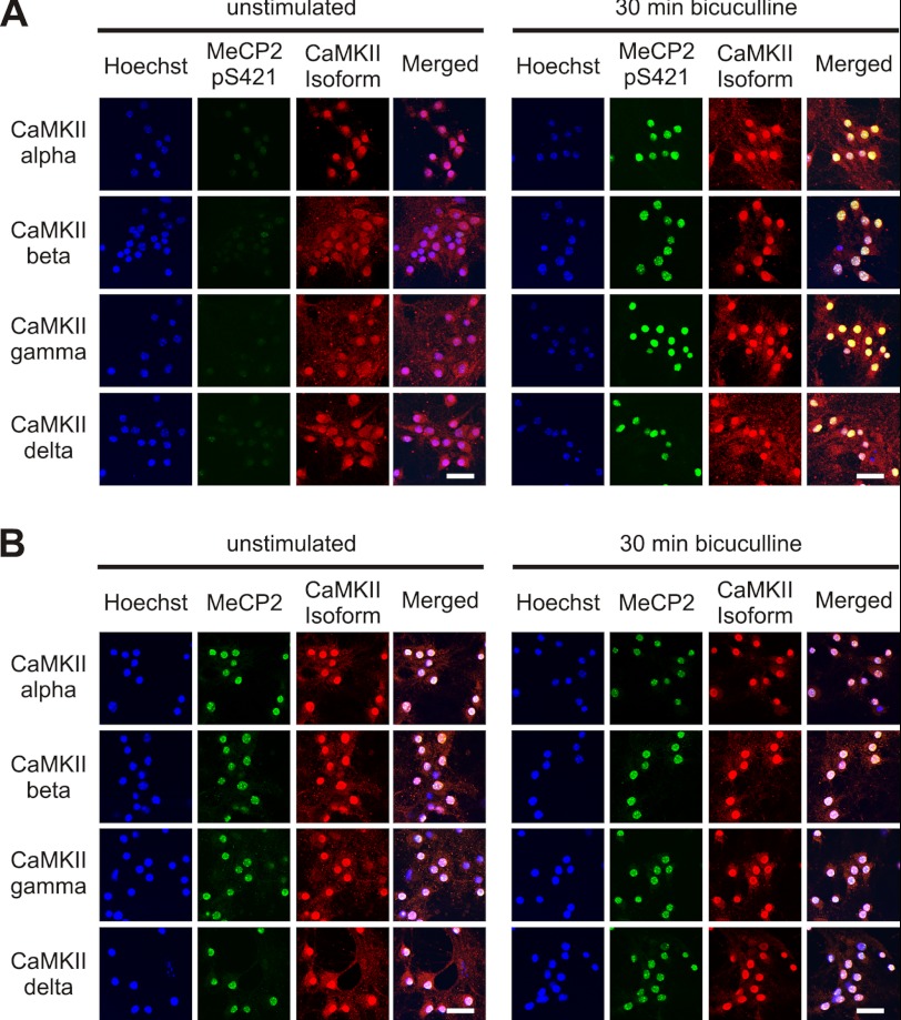
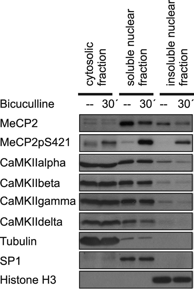
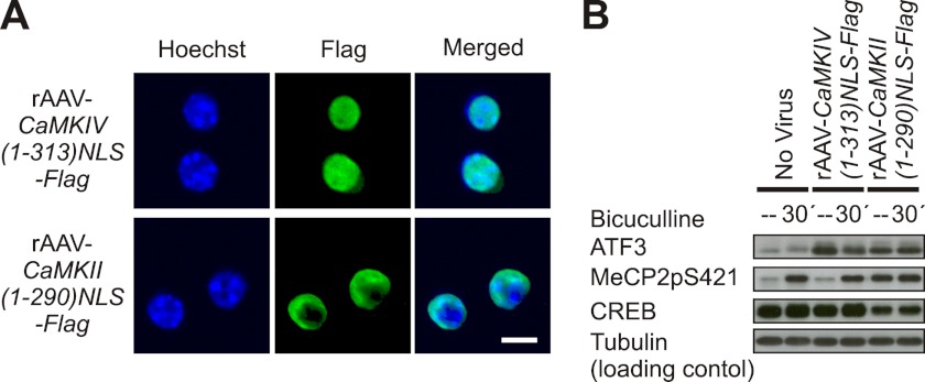
The function of MeCP2, a methylated DNA-interacting protein that may act as a global chromatin modifier, is controlled by its phosphorylation on serine 421. Here we show that in hippocampal neurons, nuclear calcium signaling controls synaptic activity-induced phosphorylation of MeCP2 on serine 421. Pharmacological inhibition of calcium/calmodulin-dependent protein (CaM)kinases blocked activity-induced MeCP2 serine 421 phosphorylation. CaM kinase II (CaMKII) but not CaMKIV, the major nuclear CaM kinase in hippocampal neurons, appeared to mediate this phosphorylation event. Biochemical subcellular fractionations and immunolocalization studies revealed that several isoforms of CaMKII (i.e. CaMKIIα, -β, -γ, and -δ) are expressed in the cytosol but are also detectable in the cell nucleus of hippocampal neurons, suggesting that nuclear CaMKII catalyzes MeCP2 serine 421 phosphorylation. Thus, in addition to the classical nuclear calcium-CaMKIV-CREB/CBP (cAMP-response element-binding protein/CREB-binding protein) pathway that regulates transcription of specific target genes, nuclear calcium may also modulate genome-wide the chromatin state in response to synaptic activity via nuclear CaMKII-MeCP2 signaling.


Figures

FIGURE 1.
FIGURE 1.
Blockade of nuclear calcium signaling. A, immunocytochemical analysis of rAAV-mediated expression of hrGFP and CaMBP4 in hippocampal neurons. Hoechst staining was used to identify nuclei. The scale bar is 20 μm. B, immunoblot analysis of rAAV-mediated expression of hrGFP (∼26 kDa; indicated by ▴) and CaMBP4-Flag (∼17 kDa; indicated by *) in hippocampal neurons. C, Fluo-3 calcium imaging (line graphs) and the corresponding quantitative analysis (bar graph) of the area under the curve of AP bursting-induced calcium transients in uninfected hippocampal neurons and in hippocampal neurons infected with the indicated rAAVs. AP bursting in hippocampal neurons was induced at the indicated time points with bicuculline (50 μm). Representative traces are shown. Measurements of individual cells are depicted in thin gray lines, and their means are shown in bold black lines. The area under the curve represents the integral of Fluo-3 signal above base line at the time of bicuculline application for a period of 200 s (n = 3). n.s., not significant. D, immunoblot analysis of ERK1/2 phosphorylation (pERK1/2) in uninfected hippocampal neurons and in hippocampal neurons infected with rAAV-hrGFP or rAAV-CaMBP4-Flag. Neurons were left unstimulated or were stimulated for 30 min or 4 h with bicuculline (50 μm). Tubulin was used as loading control. A representative of three independent experiments is shown.
FIGURE 2.
FIGURE 2.
Role of nuclear calcium in synaptic activity-induced phosphorylation of MeCP2. A, immunoblot analysis of AP bursting-induced phosphorylation of MeCP2 on serine 421 in hippocampal neurons infected with the indicated rAAVs. Expression of ATF3, MeCP2, and CREB and MeCP2 phosphorylation on serine 421 (MeCP2pS421) were analyzed in lysates of hippocampal neurons before and after induction of AP bursting with 50 μm bicuculline. Tubulin served as control. (left panel). Quantitative analysis of MeCP2pS421 level is shown in the right panel (n = 3). ***, p < 0.001. B, quantitative reverse transcription-PCR analysis of Bdnf expression from promoter IV in uninfected hippocampal neurons and in neurons infected with the indicated rAAVs. Neurons were stimulated for 4 h with 50 μm bicuculline or were left unstimulated (n = 7). C, immunoblot analysis of the effects of KN62 (10 μm) or KN93 (2 μm) on AP bursting-induced expression of ATF3 and phosphorylation of ERK1/2 (pERK1/2), CREB on serine 133 (P-CREB), and MeCP2 on serine 421 (MeCP2pS421). Hippocampal neurons pretreated for 1 h with the indicated drugs were stimulated for the indicated periods of time with bicuculline (50 μm) or were left unstimulated. D, Fluo-3 calcium imaging (line graphs) and the corresponding quantitative analysis of the area under the curve (bar graph) showing the effect of KN62 (10 μm) on AP bursting-induced calcium transients. KN62 and bicuculline were applied as in C. Representative traces are shown. Measurements of individual cells are depicted in thin gray lines, and their means are shown in bold black lines (n = 3). n.s., not significant. E, immunoblot analysis of AP bursting-induced phosphorylation of ERK1/2 (pERK1/2), CREB on serine 133 (P-CREB), and MeCP2 on serine 421 (MeCP2pS421) in hippocampal neurons with or without treatment with the indicated drugs. Pharmacological blockers were added to the cultures 1 h prior to stimulation with bicuculline (50 μm). A representative of three independent experiments is shown.
FIGURE 3.
FIGURE 3.
Role of CaMKII in MeCP2 phosphorylation. A, immunofluorescence analysis (upper panel) and immunoblot analysis (lower panel) of rAAV-mediated expression of recombinant CaMKIV(1–313)-Flag in hippocampal neurons and assessment of its effect on AP bursting-induced expression of CREB and ATF3 and MeCP2 phosphorylation on serine 421 (MeCP2pS421). CaMKIV(1–313)-Flag was detected with anti-FLAG antibody. Hoechst staining was used to identify nuclei. The scale bar is 10 μm (upper panel). Tubulin was used as loading control in the immunoblot analysis (n = 3) (lower panel). B, immunofluorescence analysis (upper panel) and immunoblot analysis (lower panel) of rAAV-mediated expression of recombinant CaMKII(1–290)-Flag expression in hippocampal neurons and assessment of its effect on AP bursting-induced expression of CREB and MeCP2 phosphorylation on serine 421 (MeCP2pS421). Hoechst staining was used to identify nuclei. The scale bar is 10 μm. Representative immunoblots from n = 3 experiments are shown.
FIGURE 4.
FIGURE 4.
Localization of CaMKII in hippocampal neurons. A, localization of MeCP2, MeCP2pS421, and the different CaMKII isoforms in the hippocampal neurons. A and B, immunocytochemical analysis of MeCP2pS421 (A), MeCP2 (B), and the α, β, γ, and δ CaMKII isoforms (A and B) in hippocampal neurons before and 30 min after induction of AP bursting using bicuculline (50 μm). Hoechst staining was used to identify nuclei. The scale bar is 20 μm.
FIGURE 5.
FIGURE 5.
Analysis of the subcellular localization of CaMKII isoforms. Immunoblot analysis of MeCP2, MeCP2pS421, and the α, β, γ, and δ isoforms of CaMKII in the cytosolic and in the soluble and insoluble nuclear fractions obtained from unstimulated hippocampal neurons and neurons 30 min after induction of AP bursting with 50 μm bicuculline. Tubulin, SP1, and histone H3 were used as controls for the different fractions of the cell lysates. A representative of three independent experiments is shown.
FIGURE 6.
FIGURE 6.
A constitutively active form of nuclear CaMKII is sufficient to cause phosphorylation of MeCP2 on serine 421. A, immunofluorescence analysis of rAAV-mediated expression of CaMKIV(1–313)NLS-Flag and CaMKII(1–290)NLS-Flag. Hoechst was used to identify nuclei. The scale bar is 10 μm. B, immunoblot analysis of MeCP2 phosphorylation on serine 421 (MeCP2pS421) and the expression of CREB and ATF3 in hippocampal neurons infected with the indicated rAAVs before and 30 min after induction of AP bursting using 50 μm bicuculline. Tubulin served as loading control. A representative immunoblot from n = 3 experiments is shown.

Similar articles

Cited by

References

    1. Hendrich B., Bird A. (1998) Identification and characterization of a family of mammalian methyl-CpG-binding proteins. Mol. Cell Biol. 18, 6538–6547 - PMC - PubMed
    1. Fuks F., Hurd P. J., Wolf D., Nan X., Bird A. P., Kouzarides T. (2003) The methyl-CpG-binding protein MeCP2 links DNA methylation to histone methylation. J. Biol. Chem. 278, 4035–4040 - PubMed
    1. Chahrour M., Jung S. Y., Shaw C., Zhou X., Wong S. T., Qin J., Zoghbi H. Y. (2008) MeCP2, a key contributor to neurological disease, activates and represses transcription. Science 320, 1224–1229 - PMC - PubMed
    1. Chen W. G., Chang Q., Lin Y., Meissner A., West A. E., Griffith E. C., Jaenisch R., Greenberg M. E. (2003) Derepression of BDNF transcription involves calcium-dependent phosphorylation of MeCP2. Science 302, 885–889 - PubMed
    1. Shahbazian M. D., Antalffy B., Armstrong D. L., Zoghbi H. Y. (2002) Insight into Rett syndrome: MeCP2 levels display tissue- and cell-specific differences and correlate with neuronal maturation. Hum. Mol. Genet. 11, 115–124 - PubMed

Publication types

MeSH terms

Substances

LinkOut - more resources