. 2012 Sep 7;287(37):31223-32.
doi: 10.1074/jbc.M111.314583. Epub 2012 Jul 20.

Endoplasmic reticulum stress induces a caspase-dependent N-terminal cleavage of RBX1 protein in B cells

Affiliations

Endoplasmic reticulum stress induces a caspase-dependent N-terminal cleavage of RBX1 protein in B cells

Shimon Shteingart et al. J Biol Chem. .

Abstract

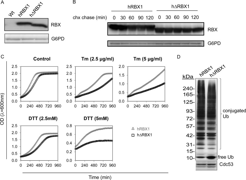
Endoplasmic reticulum (ER) stress develops when the ER is overloaded with too many proteins to fold. This elicits a signaling pathway called the unfolded protein response. The unfolded protein response is physiologically required for the terminal development of B cells into antibody-secreting plasma cells. Ring Box Protein 1 (RBX1) is a 14-kDa protein necessary for ubiquitin ligation activity of the multimeric cullin ring ubiquitin ligases (CRLs). As RBX1 is shared by a large number of CRLs, alterations in its activity may lead to global changes in protein stability. We discovered that RBX1 is cleaved in the course of LPS-induced plasma cell differentiation and in multiple myeloma cell lines upon induction of pharmacological ER stress. The cleavage is executed by several caspase proteases that cleave RBX1 eight amino acids from the N terminus. To address the possible implication of RBX1 cleavage for CRL activity, we replaced the endogenous RBX1 homolog of the yeast Saccharomyces cerevisiae, Roc1, with the wild type or the N-terminal Δ8 mutant human RBX1. We show that yeast expressing the cleaved RBX1 are hypersensitive to ER stress and are impaired in CRL-mediated ubiquitination and degradation. We propose a model by which N-terminal cleavage of RBX1 impairs its activity and promotes susceptibility to ER stress induction.


Figures

FIGURE 1.
FIGURE 1.
Rbx1 is cleaved during B cell differentiation and ER stress induction. A, B cells were purified from spleens of WT or Bcl2 Tg mice and incubated for up to 4 days with LPS. Total cell lysates were prepared, analyzed on 15% SDS-PAGE, and blotted with anti-RBX1 antibody. B, i.29μ+ cells were incubated with LPS, and samples were analyzed as in A. C, RPMI8226 cells were treated overnight with the indicated compound. Total cell extract was prepared and analyzed as in A. D, RPMI8226 cells were treated overnight with 2.5 μg/ml and subjected to CHX treatment for up to 4 h. Samples of equal numbers of cells were withdrawn and analyzed for RBX1 as in A. Shown are typical experiments of at least three repetitions.
FIGURE 2.
FIGURE 2.
RBX1 is cleaved by caspases after Asp-8. A, RPMI8226 cells were treated with 2.5 μg/ml Tg, and in the presence of the indicated caspase inhibitors (50 μm), RBX1 cleavage was assessed by immunoblotting of cell lysates with anti-RBX1. B, 293T cells were lysed in protease buffer. 35 μg of total proteins incubated each of the caspases individually, and reaction was stopped by boiling in sample buffer. The reaction was then analyzed by Western blotting with anti-RBX1 antibody. C, splenic B cells were purified from heterozygous or caspase-1 KO mice and incubated for 3 days with LPS. 2.5 μg/ml Tg was added at the last 24 h where indicated. Cell lysates were separated on 15% SDS-PAGE and blotted with anti-RBX1 antibody. D, pcDNA3.1 vector expressing the indicated RBX1 mutant was transfected into 293T cells, and following 48 h cell lysates were analyzed by Western blotting with anti-RBX1. E, recombinant GST-RBX1 with Asp residue substitutions at position 6 and/or 8 were expressed in BL21 E. coli and incubated with rCaspase-1. Cleavage was detected using Western blotting with anti-RBX1. F, pcDNA3.1 that encodes V5(D2E)-tagged Rbx1 or its AVA mutant was electroporated into RPMI8226 cells and treated or not overnight with Tg. Total cell extracts were immunoblotted for V5 and Rbx1.
FIGURE 3.
FIGURE 3.
ΔhRBX1 binds to Cul1. ΔRBX1 is stable upon transfection with pcDNA3.1 that encodes ΔRBX1, followed by immunoblotting in the presence of CHX for 4 h (A). Plasmids expressing GST-cullin1 and RBX1 or ΔhRBX1 were co-transfected into 293T cells. Cells were harvested after 48 h. After mild lysis, whole cell extracts (WCL) were pulled down with GST beads. Samples were separated on 10 and 16% SDS-PAGE and blotted with anti-Cul1 and anti-RBX1 antibodies, respectively (B).
FIGURE 4.
FIGURE 4.
Yeast expressing hΔRBX1 are hypersensitive to ER stress. Yeasts deleted of their endogenous homolog of RBX1 and transgenic for either RBX1 or ΔhRBX1 were lysed and immunoblotted with anti-hRBX1 antibody, where wild type yeast served as a negative control (A). Both RBX1 forms were stable for 2 h during CHX chase (B). Growth tests were performed using a Tecan plate reader in a flat-bottomed 96-well plate. Absorbance measurements were taken in 15-min intervals. Chemicals were added to the wells at t = 0 (C). Shown is immunoblot analysis for ubiquitin and Cdc53 from hRBX1 or ΔhRBX1 expressing strains, harvested at the logarithmic growth phase at equal absorbance. Densitometry analysis of four repeats shows ∼15% reduction in ubiquitination in the ΔhRBX1 strain and a similar increase at the level of free ubiquitin (D).
FIGURE 5.
FIGURE 5.
ER stress causes higher UPR induction in ΔhRBX1-expressing cells. Yeast cells were transformed with a plasmid encoding β-gal, under the control of a UPRE promoter. Samples were collected at the indicated times after ER stress induction by Tm (2 μg/ml). β-Gal activity was assayed and normalized to the sample total protein concentration (A). Yeast were transformed with HA-tagged HAC1p, and CHX (0.5 mg/ml) was added to mid-log phase culture. Samples were collected for immunoblotting at indicated time points. Hac1p levels were quantified and normalized against glucose-6-phosphate dehydrogenase (G6PD) levels (B). Immunoblotting to hRBX1 was performed to confirm strain identity.

Similar articles

Cited by

References

    1. Ron D., Walter P. (2007) Signal integration in the endoplasmic reticulum unfolded protein response. Nat. Rev. Mol. Cell Biol. 8, 519–529 - PubMed
    1. Travers K. J., Patil C. K., Wodicka L., Lockhart D. J., Weissman J. S., Walter P. (2000) Functional and genomic analyses reveal an essential coordination between the unfolded protein response and ER-associated degradation. Cell 101, 249–258 - PubMed
    1. Yamamoto K., Sato T., Matsui T., Sato M., Okada T., Yoshida H., Harada A., Mori K. (2007) Transcriptional induction of mammalian ER quality control proteins is mediated by single or combined action of ATF6α and XBP1. Dev. Cell 13, 365–376 - PubMed
    1. Zhang K., Wong H. N., Song B., Miller C. N., Scheuner D., Kaufman R. J. (2005) The unfolded protein response sensor IRE1α is required at two distinct steps in B cell lymphopoiesis. J. Clin. Invest. 115, 268–281 - PMC - PubMed
    1. Reimold A. M., Iwakoshi N. N., Manis J., Vallabhajosyula P., Szomolanyi-Tsuda E., Gravallese E. M., Friend D., Grusby M. J., Alt F., Glimcher L. H. (2001) Plasma cell differentiation requires the transcription factor XBP-1. Nature 412, 300–307 - PubMed

Publication types