

Review
doi: 10.1074/jbc.R112.349068.
Epub 2012 Jul 20.
Ryanodine receptors: structure and function
Affiliations
- PMID: 22822064
- PMCID: PMC3442496
- DOI: 10.1074/jbc.R112.349068
Item in Clipboard
Review
Ryanodine receptors: structure and function
J Biol Chem.
.
Abstract
Ryanodine receptors (RyRs) are huge ion channels that are responsible for the release of Ca(2+) from the sarco/endoplasmic reticulum. RyRs form homotetramers with a mushroom-like shape, consisting of a large cytoplasmic head and transmembrane stalk. Ca(2+) is a major physiological ligand that triggers opening of RyRs, but a plethora of modulatory proteins and small molecules in the cytoplasm and sarco/endoplasmic reticulum lumen have been recognized. Over 300 mutations in RyRs are associated with severe skeletal muscle disorders or triggered cardiac arrhythmias. With the advent of high-resolution structures of individual domains, many of these can be mapped onto the three-dimensional structure.
Figures
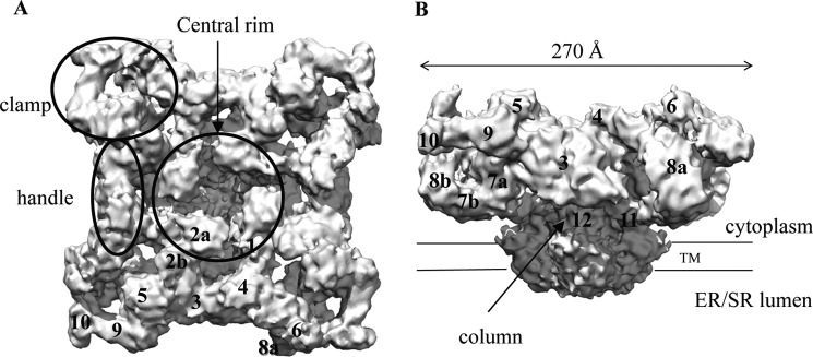
Overall structure. Shown is a cryo-EM reconstruction of RyR1 at 9.6 Å (Electron Microscopy Data Bank entry 1275) (24). A, top view from the cytoplasm, looking toward the SR/ER. B, side view showing the large cytoplasmic head. Labels show the structural elements and the numbered subregions. TM, transmembrane domain.
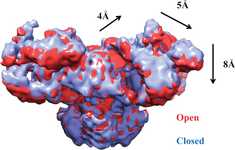
Allosteric movements. Shown is the superposition of RyR1 cryo-EM maps in the closed (blue) and open (red) states (Electron Microscopy Data Bank entries 1606 and 1607) (22). The arrows and distances show the overall motions in the cytoplasmic region as the RyR opens.
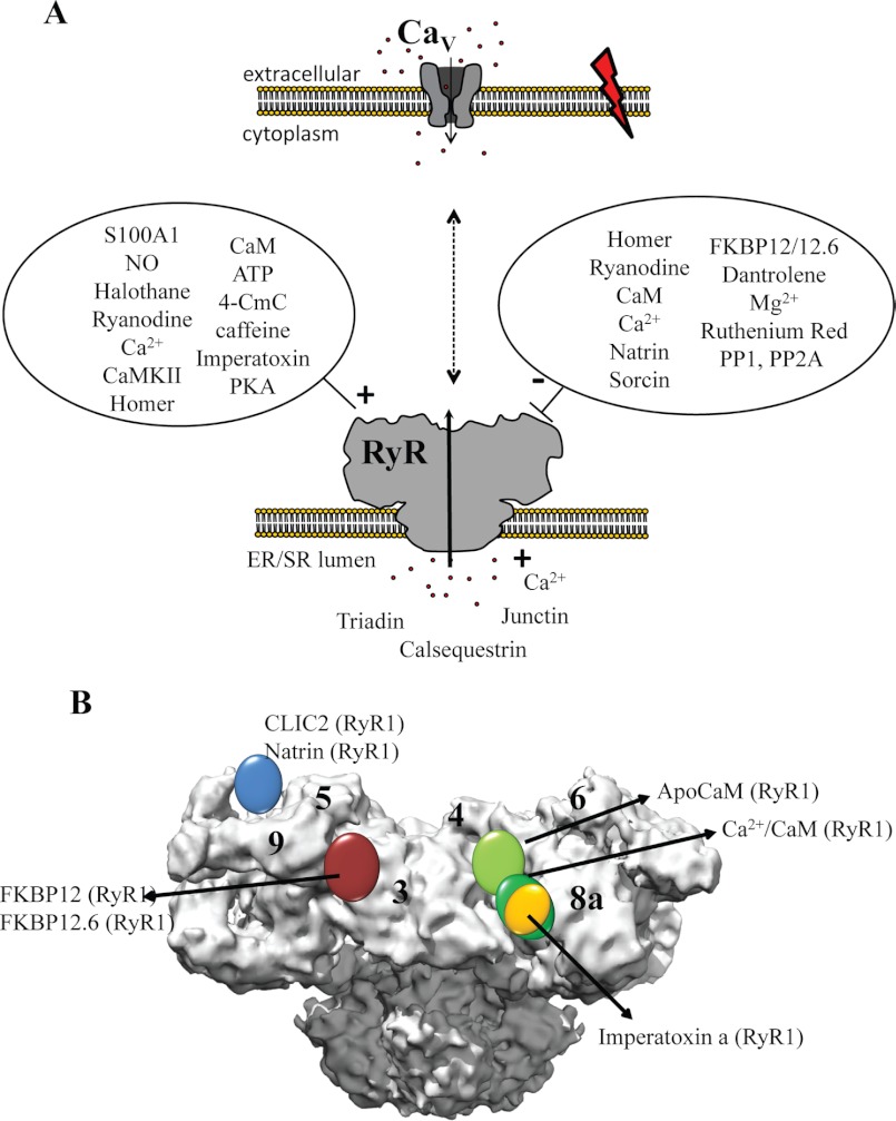
Binding partners and ligands.
A, schematic overview of the RyR and voltage-gated calcium channel (CaV), present in two different membranes, along with several binding partners in the cytoplasmic and luminal areas. 4-CmC, 4-chloro-m-cresol. B, locations of several protein-binding partners based on difference cryo-EM.
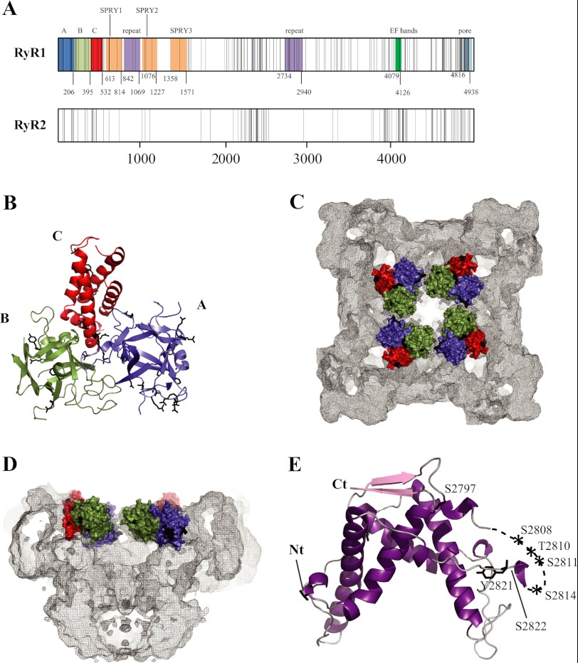
Disease hot spots.
A, linear view of the RyR sequences, with each vertical line representing a disease mutation. The shaded areas indicate experimentally verified (domains A–C) and some key predicted domains. Another truncated repeat may be present between SPRY2 and SPRY3 (not shown). The domain boundaries are numbered according to the RyR1 sequence. Except for domains A–C, the numbering is only a prediction and may vary (Ref. and the Phyre2 server, Structural Informatics Group, Imperial College London). The predicted pore-forming area is based on homology to the pore-forming region of a bacterial voltage-gated sodium channel (Protein Data Bank code 3RVZ). B, crystal structure of the RyR1 ABC region (Protein Data Bank code 2XOA) (93). Disease mutations are shown as black sticks. Mutations in flexible loops have been omitted for clarity. C and D, location of the RyR1 ABC domains in the RyR1 cryo-EM map. The domains are shown in surface representation. Black patches correspond to the locations of disease mutations. Shown are a view from the cytoplasm (C) and a side view with one-half omitted (D). E, crystal structure of the RyR2 phosphorylation domain. The labels indicate phosphorylation target sites within the phosphorylation loop.
References
-
- Rogers E. F., Koniuszy F. R., Shavel J., Jr., Folkersand K. (1948) Plant insecticides. I. Ryanodine, a new alkaloid from Ryania speciosa Vahl. J. Am. Chem. Soc. 70, 3086–3088 - PubMed
-
- Lai F. A., Erickson H. P., Rousseau E., Liu Q. Y., Meissner G. (1988) Purification and reconstitution of the calcium release channel from skeletal muscle. Nature 331, 315–319 - PubMed
-
- Inui M., Saito A., Fleischer S. (1987) Purification of the ryanodine receptor and identity with feet structures of junctional terminal cisternae of sarcoplasmic reticulum from fast skeletal muscle. J. Biol. Chem. 262, 1740–1747 - PubMed
-
- Meissner G. (1986) Ryanodine activation and inhibition of the Ca2+ release channel of sarcoplasmic reticulum. J. Biol. Chem. 261, 6300–6306 - PubMed
Publication types
MeSH terms
Substances
Grants and funding
LinkOut - more resources
Full Text Sources
Other Literature Sources
Molecular Biology Databases
Miscellaneous
