Review
. 2012 Sep 14;287(38):31633-40.
doi: 10.1074/jbc.R112.349464. Epub 2012 Jul 20.

Cyclic ADP-ribose and nicotinic acid adenine dinucleotide phosphate (NAADP) as messengers for calcium mobilization

Affiliations
Review

Cyclic ADP-ribose and nicotinic acid adenine dinucleotide phosphate (NAADP) as messengers for calcium mobilization

Hon Cheung Lee. J Biol Chem. .

Abstract

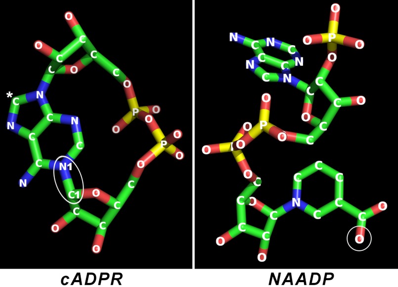
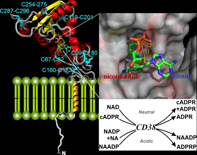
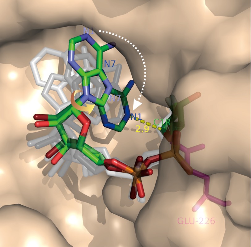
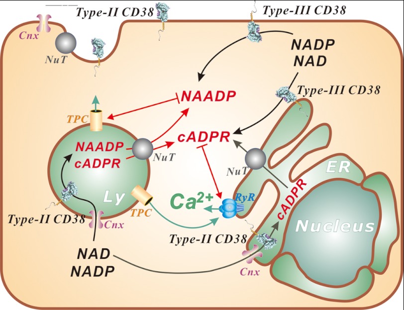
Cyclic ADP-ribose and nicotinic acid adenine dinucleotide phosphate were discovered >2 decades ago. That they are second messengers for mobilizing Ca(2+) stores has since been firmly established. Separate stores and distinct Ca(2+) channels are targeted, with cyclic ADP-ribose acting on the ryanodine receptors in the endoplasmic reticulum, whereas nicotinic acid adenine dinucleotide phosphate mobilizes the endolysosomes via the two-pore channels. Despite the structural and functional differences, both messengers are synthesized by a ubiquitous enzyme, CD38, whose crystal structure and catalytic mechanism have now been well elucidated. How this novel signaling enzyme is regulated remains largely unknown and is the focus of this minireview.


Figures

FIGURE 1.
FIGURE 1.
Crystal structures of cADPR and NAADP. The structure of cADPR was obtained from crystals of its free acid, and that of NAADP was from the crystals of the complex of NAADP and CD38. The cyclization site in cADPR is indicated by an ellipse. The C8 of cADPR is indicated by a white asterisk. Attachment of a bromo (8-bromo-cADPR) or an amino (8-NH2-cADPR) group at this position converts the compound to a specific antagonist of cADPR. The structure of NAADP is identical to that of its parent NADP, except that the amide nitrogen of the nicotinamide group of NADP is changed to oxygen as indicated by the circle. Blue, nitrogen; red, oxygen; yellow, phosphorus; green, carbon.
FIGURE 2.
FIGURE 2.
Crystal structure of CD38. The secondary structure of the C-terminal domain of CD38 is shown, with helices in red, β-sheets in yellow, and coils in gray. The disulfides are labeled and colored cyan. The transmembrane segment is modeled as a gold helix, whereas the N-terminal tail as a random coil. A molecule of NAD is bound at the active site near the middle of the C-terminal domain and is rendered in stick configuration. Red, oxygen; orange, phosphorus; green, carbon; blue, nitrogen. The upper inset shows the active site in surface view. The nicotinamide end of the bound NAD enters the site first, whereas the adenine end is positioned toward the opening of the site. The lower inset lists the multiple reactions catalyzed by CD38. The synthesis and hydrolysis of cADPR occur at neutral pH, whereas those of NAADP occur at acidic pH. NA, nicotinic acid, ADPRP, ADP-ribose phosphate.
FIGURE 3.
FIGURE 3.
Imaging the NAD cyclization by CD38. A mutant of CD38 with Glu-146 changed to alanine (described in text) was co-crystallized with a close analog of NAD, 2′-deoxy-2′-fluoroarabinoside NAD, which forms a covalent linkage between its ribosyl anomeric carbon (C1R) and the catalytic residue Glu-226. Six molecules of the complex are found in each of the asymmetric crystal units, each containing a covalent intermediate in a different conformation. The conformations relate to each other by single-bond rotation, indicating that they represent substates of the cyclization process. Ordering the conformations shows that the cyclization requires a 180° rotation of the adenine ring around its glycosidic bond (yellow arrow), such that the N1 of the adenine is put into close proximity to the ribosyl anomeric carbon (2.9 Å; yellow dashed line), the site of cyclization. Rendered in colored sticks are the starting state, where the adenine ring is farthest from the ribosyl anomeric carbon, and the ending state, where the two are closest. The substates representing various degrees of single-bond rotation of the intermediate between the starting and ending states are colored in shades of gray. Red, oxygen; orange, phosphorus; green, carbon; blue, nitrogen.
FIGURE 4.
FIGURE 4.
Emerging mechanisms of regulating the signaling function of CD38. NAADP activates the TPCs in the endolysosomes (Ly), whereas cADPR targets the ryanodine receptors (RyR) in the ER. Ca2+ released by NAADP from the endolysosomal stores can be amplified Ca2+-induced Ca2+ release from ER stores. This process is potentiated by cADPR through its sensitization of the ryanodine receptors to Ca2+. CD38 can be coexpressed with both type II and III membrane orientations. Type II CD38 has its catalytic domain facing either outside of the cell or inside the lumen of organelles. Regulation of type II CD38 is proposed to be through substrate limitation, as both NAD and NADP are in the cytosol. Their transport into the organelles is mediated by connexin-43 (Cnx), whereas the products cADPR and NAADP, produced in the lumen, can likewise be transported out to the cytosol by the nucleoside transporters (NuT). Both nucleoside transporters and connexin-43 are also present on the cell surface to serve a similar function for signaling by the ecto-expressed CD38. Type III CD38 is amenable to many common regulation mechanisms, such as phosphorylation.

Similar articles

Cited by

References

    1. Streb H., Irvine R. F., Berridge M. J., Schulz I. (1983) Release of Ca2+ from a non-mitochondrial intracellular store in pancreatic acinar cells by inositol 1,4,5-trisphosphate. Nature 306, 67–69 - PubMed
    1. Lee H. C., Aarhus R. (1995) A derivative of NADP mobilizes calcium stores insensitive to inositol trisphosphate and cyclic ADP-ribose. J. Biol. Chem. 270, 2152–2157 - PubMed
    1. Lee H. C., Aarhus R., Levitt D. (1994) The crystal structure of cyclic ADP-ribose. Nat. Struct. Biol. 1, 143–144 - PubMed
    1. Lee H. C., Walseth T. F., Bratt G. T., Hayes R. N., Clapper D. L. (1989) Structural determination of a cyclic metabolite of NAD+ with intracellular Ca2+-mobilizing activity. J. Biol. Chem. 264, 1608–1615 - PubMed
    1. Wu Y., Kuzma J., Maréchal E., Graeff R., Lee H. C., Foster R., Chua N. H. (1997) Abscisic acid signaling through cyclic ADP-ribose in plants. Science 278, 2126–2130 - PubMed

Publication types