Review
. 2012 Sep 14;287(38):31641-9.
doi: 10.1074/jbc.R112.353573. Epub 2012 Jul 20.

Ca2+ regulation of ion transport in the Na+/Ca2+ exchanger

Affiliations
Review

Ca2+ regulation of ion transport in the Na+/Ca2+ exchanger

Mark Hilge. J Biol Chem. .

Abstract

The binding of Ca(2+) to two adjacent Ca(2+)-binding domains, CBD1 and CBD2, regulates ion transport in the Na(+)/Ca(2+) exchanger. As sensors for intracellular Ca(2+), the CBDs form electrostatic switches that induce the conformational changes required to initiate and sustain Na(+)/Ca(2+) exchange. Depending on the presence of a few key residues in the Ca(2+)-binding sites, zero to four Ca(2+) ions can bind with affinities between 0.1 to 20 μm. Importantly, variability in CBD2 as a consequence of alternative splicing modulates not only the number and affinities of the Ca(2+)-binding sites in CBD2 but also the Ca(2+) affinities in CBD1.


Figures
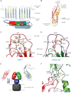
FIGURE 1.
FIGURE 1.
Structural basis and organization of the two Ca2+ sensors (CBD1 and CBD2) in NCX. A, updated topology model of NCX consisting of a transmembrane domain and an ∼500-residue-long cytosolic loop that harbors the two Ca2+-binding domains (CBD1 and CBD2). The variable region in CBD2 as a consequence of alternative splicing (AS) is shown in violet. The conserved α-repeat regions that define the cation/Ca2+ exchanger superfamily are colored yellow. B, ribbon diagram displaying the β-sandwich architecture of Ca2+-bound CBD1. The structures of the CBDs redefined the previously described Calx-β motif by adding strands A and G (yellow). C and D, detailed views of the CBD1 (PDB code 2DPK) and CBD2 (code 2QVM) Ca2+-binding sites in the Ca2+-bound form. E, hypothetical model of NCX consisting of four domains: transmembrane domain (residues 1–217 and 727–903; gray), the CLD (residues 218–370 and 651–726; blue), CBD1 (residues 371–500; red), and CBD2 (residues 501–650; green), with the numbering based on the canine NCX1 AD splice variant (NCX1.4). The ribbon diagram of CBD2 (upper right) displays the variable region encoded by the mutually exclusive exons A or B (blue) and the small cassette exons (yellow) at the opposite side of the CBD2 Ca2+-binding sites. F, interface of CBD1 and CBD2, displaying the crucial salt bridges and hydrogen bonds in the presence of Ca2+. Residues in parentheses refer to the numbering scheme of canine NCX1.
FIGURE 2.
FIGURE 2.
Structure of the NCX homolog from M. jannaschii and a hypothetical model for intact NCX1. A, ribbon diagram displaying symmetry-related helices 1–5 (light gray) and 6–10 (dark gray). The inset shows the detailed coordination of three Na+ ions and one Ca2+ ion. B, summary of the available structural data assembled in a hypothetical model for intact NCX1. Residues for which no structural data are available are indicated. The orientation of the domains with respect to each other is arbitrary, as the linker regions between the CLD and the transmembrane domain, as well as the CBDs, are missing. The model for the transmembrane domain of NCX1 is based on the structure of NCX_Mj (PDB code 3V5U), whereas the homology model of the CLD is derived from the structure of α-catenin (code 1H6G).
FIGURE 3.
FIGURE 3.
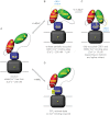
Hypothetical dual electrostatic switch mechanism of Ca2+ regulation in NCX. A, inactive Ca2+-free NCX in extended conformation. B, submicromolar Ca2+ concentrations induce a conformational change via the electrostatic switch in CBD1 that results in a compaction of the CBDs and that probably reduces tension on the linker regions to the CLD. C, binding of Ca2+ to CBD2 allows sustained Na+/Ca2+ exchange and removes counteracting Na+-dependent inactivation. Whether Ca2+ binding to CBD2 induces yet another conformational change or just promotes an interaction with the CLD remains elusive. D, depiction of the PIP2-activated state of NCX that is independent of Ca2+ binding the CBDs.

Similar articles

Cited by

References

    1. Blaustein M. P., Lederer W. J. (1999) Sodium/calcium exchange: its physiological implications. Physiol. Rev. 79, 763–854 - PubMed
    1. Philipson K. D., Nicoll D. A. (2000) Sodium/calcium exchange: a molecular perspective. Annu. Rev. Physiol. 62, 111–133 - PubMed
    1. Annunziato L., Pignataro G., Di Renzo G. F. (2004) Pharmacology of brain Na+/Ca2+ exchanger: from molecular biology to therapeutic perspectives. Pharmacol. Rev. 56, 633–654 - PubMed
    1. Lytton J. (2007) Na+/Ca2+ exchangers: three mammalian gene families control Ca2+ transport. Biochem. J. 406, 365–382 - PubMed
    1. Brini M., Carafoli E. (2011) The plasma membrane Ca2+-ATPase and the plasma membrane sodium/calcium exchanger cooperate in the regulation of cell calcium. Cold Spring Harb. Perspect. Biol. 3, a004168. - PMC - PubMed

Publication types

LinkOut - more resources