. 2012 Sep 7;287(37):31515-26.
doi: 10.1074/jbc.M112.396895. Epub 2012 Jul 22.

Identifying key juxtamembrane interactions in cell membranes using AraC-based transcriptional reporter assay (AraTM)

Affiliations

Identifying key juxtamembrane interactions in cell membranes using AraC-based transcriptional reporter assay (AraTM)

Pin-Chuan Su et al. J Biol Chem. .

Abstract

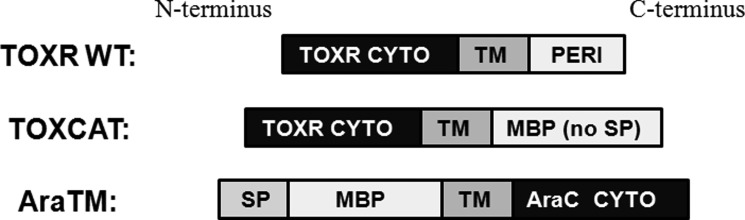
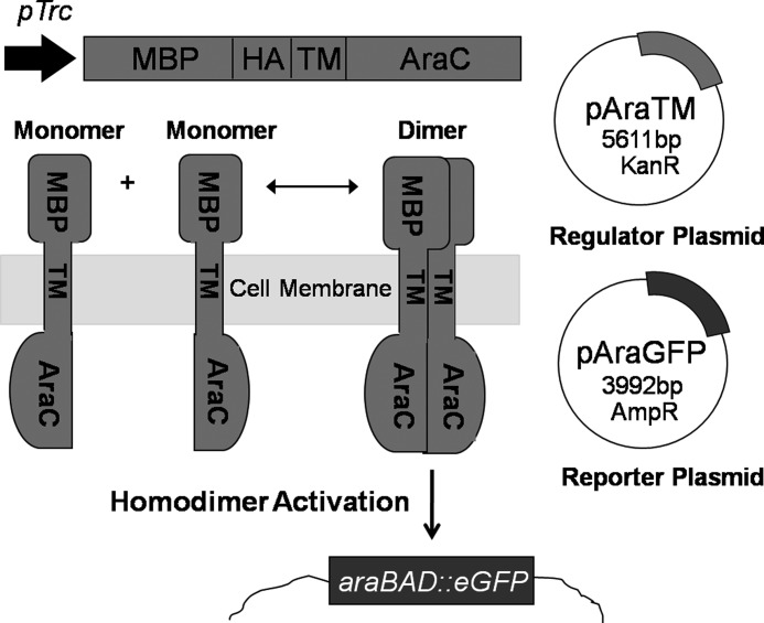
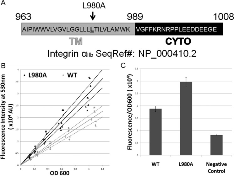
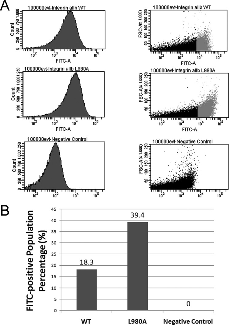
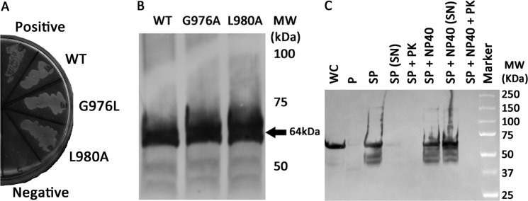
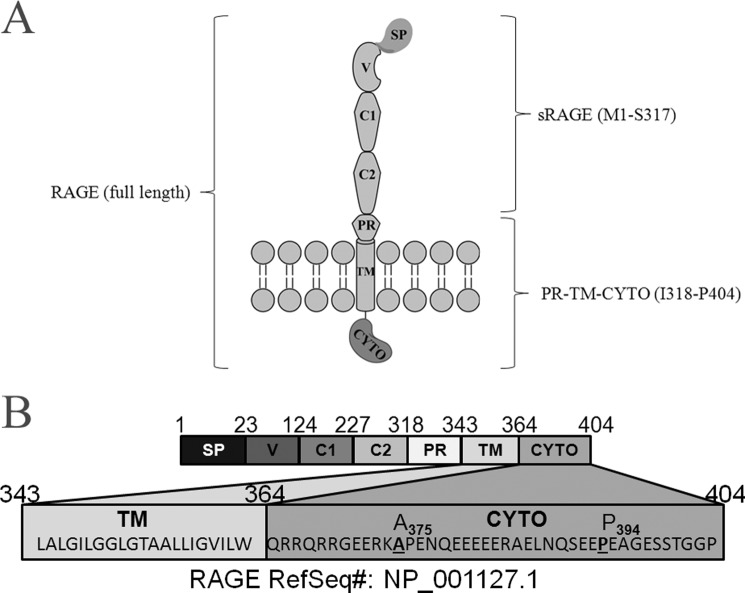
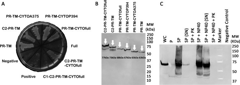
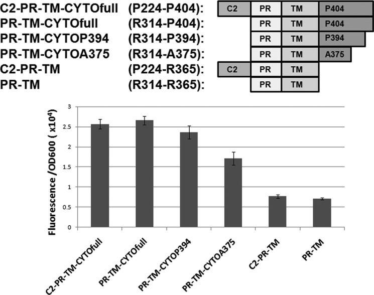
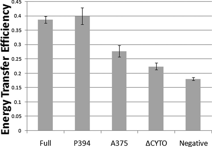
Dimerization is a key regulatory mechanism in activation of transmembrane (TM) receptors during signal transduction. This process involves a coordinated interplay between extracellular (EX), TM, and cytoplasmic (CYTO) regions to form a specific interface required for both ligand binding and intracellular signaling to occur. While several transcriptional activator-based methods exist for investigating TM interactions in bacterial membranes, expression of TM chimera in these methods occurs in a reverse orientation, and are limited to only TM domains for proper membrane trafficking and integration. We therefore developed a new, AraC-based transcriptional reporter assay (AraTM) that expresses EX-TM-CYTO chimera in their native orientation, thereby enabling membrane trafficking to occur independent of the TM chimera used as well as permitting analysis of EX-TM-CYTO interactions in biological membranes. Using integrin α(IIb) TM-CYTO as a model, we observe a large increase in homodimerization for the constitutively active TM mutant L980A relative to wild-type in the TM-CYTO construct (A963-E1008). We also characterized the receptor for advanced glycation endproducts (RAGE), whose homooligomeric state is critical in ligand recognition, and find the specific juxtamembrane region within the CYTO (A375-P394) mediates homodimerization, and is dominant over effects observed when the extracellular C2 domain is included. Furthermore, we find good agreement between our AraTM measurements in bacterial membranes and BRET measurements made on corresponding RAGE constructs expressed in transfected HEK293 cells. Overall, the AraTM assay provides a new approach to identify specific interactions between receptor EX-TM-CYTO domains in biological membranes that are important in regulation of signal transduction.


Figures

FIGURE 1.
FIGURE 1.
Organization of AraC and ToxR Fusions Used in TM Interaction Assays. In ToxR-based assays (–14), constructs are configured with ToxR as an N-terminal fusion to a TM domain of interest, with mature MBP lacking its signal peptide (SP) fused at the C terminus. ToxR is a type II integral membrane protein (18), and therefore the TM domain of interest acts both as a signal peptide to direct membrane trafficking as well as membrane integration with the TM domain in a reverse orientation. In the AraTM assay, the C-terminal orientation of the DNA-binding domain within AraC enables constructs to be expressed with a N-terminal MBP fusion, which includes its native signal peptide to direct membrane integration. Thus, AraTM constructs are expressed in their native orientation, and membrane integration is decoupled from the specific sequence fused to MBP and AraC.
FIGURE 2.
FIGURE 2.
Overview of AraTM assay. Chimeric proteins containing N-terminal MBP and C-terminal AraC domains fused with an in-frame receptor fragment are expressed by the regulator plasmid (pAraTM; kanamycin resistant). Once expressed, MBP directs expression of the chimera to the inner membrane of E. coli. Homodimerization brings AraC transcriptional factors in close proximity, enabling binding to the PBAD promoter on the reporter plasmid (pAraGFP; ampicillin resistance) and activating transcription of the reporter gene GFP.
FIGURE 3.
FIGURE 3.
AraTM assay results for wild-type integrin αIIb TM-CYTO and its mutations. A, integrin αIIb TM-CYTO sequence subcloned into pAraTM. The L980A mutation is also highlighted. B, fluorescence intensity at 530 nm of wild-type integrin αIIb TM-CYTO and mutant L980A from serial dilutions of bacterial cultures plotted against the corresponding cell density (OD600). Solid lines represent the best-fit line through the experimental data, and the dotted lines represent the upper and lower bound of the 95% confidence interval for each data set. C, average slopes from fluorescence intensity versus OD600 for each construct are compared from eight independent replicates, with error bars representing standard error.
FIGURE 4.
FIGURE 4.
Population distribution of GFP-positive cells for various integrin αIIb AraTM constructs by flow cytometry. A, compared with wild-type, the mutant L980A shows an increase in the % of total cell population (100,000 events) that is GFP-positive as well as a shift in the overall cell population measured in terms of FSC to a higher average GFP signal, which is in good agreement with whole-cell fluorescence measurements from cell suspension (Fig. 3). B, integrin αIIb L980A mutant shows a 2-fold increase in the GFP-positive population as compared with wild-type.
FIGURE 5.
FIGURE 5.
The integrin αIIb TM-CYTO chimera express at similar levels and are properly integrated into the inner membrane of E. coli. A, AraTM chimeras containing wild-type and mutant integrin αIIb TM-CYTO expressed in MalE-deficient MM39 cells were streaked on a 0.4% maltose M9 plate and incubated for 48 h at 37 °C. Each construct is properly integrated into the inner membrane of E. coli, as indicated by robust growth on the 0.4% maltose M9 plates similar to the positive control (pTrcRSF containing MBP-AraC chimera). As expected, no growth is observed on the negative control (AraCY). B, wild-type and mutant integrin αIIb TM-CYTO chimera were expressed at equal levels as determined by immunoblotting with HRP-conjugated anti-MBP antibody, and the observed chimera MWs were consistent with the expected MWs. C, periplasts and spheroplasts were prepared for mutant integrin αIIb L980A TM-CYTO, treated with and without Nonidet P-40 (1% v/v) and proteinase K (50 μg/ml), and blotted against anti-MBP antibody (WC: whole cell, P: periplast, SP: spheroplast, SN: supernatant, PK: proteinase K, and Nonidet P-40: Detergent Nonidet P-40). No chimera is detected in intact spheroplasts treated with proteinase K (SP + PK) nor in the periplasmic fraction of the cell, consistent with the expected periplasmic orientation and membrane integration of the MBP fusion.
FIGURE 6.
FIGURE 6.
Monitoring expression of AraTM chimera in cell culture. Cells expressing integrin αIIb L980A chimera were collected hourly to monitor expression level and degradation products by immunoblotting with an anti-MBP antibody. Chimera expression began 3 h post-induction at 37 °C and remained constant overnight.
FIGURE 7.
FIGURE 7.
RAGE. A, graphic illustration of domain structure in the full-length receptor, including the TM and CYTO regions. B, annotation of amino acid sequences in full-length RAGE corresponding to specific domains within the receptor. Sequences for the TM and CYTO region of RAGE are given, including positions (A375, P394) for specific truncations in the CYTO region (SP: signal peptide, V: V domain, C1: C1 domain, C2: C2 domain, PR: proximal domain).
FIGURE 8.
FIGURE 8.
Expression of RAGE constructs in AraTM assay. A, RAGE C2-PR-TM-CYTOfull AraTM chimera as well as additional truncations of RAGE C2-PR-TM-CYTOfull are able to complement growth on maltose M9 minimal plates. B, cells expressing RAGE chimera were expressed at similar levels and the chimera MWs are consistent with the expected MWs as determined by immunoblotting from whole-cell lysates with anti-MBP antibody. C, periplasts and spheroplasts of the RAGE PR-TM-CYTOfull chimera were prepared, treated with/without Nonidet P-40 (1% v/v) and proteinase K (50 μg/ml), and blotted against anti-MBP antibody (WC: whole cell, P: periplast, SP: spheroplast, SN: supernatant, PK: proteinase K, and Nonidet P-40: detergent Nonidet P-40). No chimera is detected in intact spheroplasts treated with proteinase K (SP + PK) nor in the periplasmic fraction of the cell, consistent with the expected periplasmic orientation and membrane integration of the MBP-RAGE-AraC fusion.
FIGURE 9.
FIGURE 9.
Cytoplasmic truncations of RAGE reduce homodimerization in cell membranes. Removing the last 10 amino acids in the cytosolic domain of RAGE (PR-TM-CTYOP394) had minimal affect on RAGE dimerization, whereas removal of the central domain (PR-TM-CYTOA375) reduced dimerization, but not to background levels observed in the cytoplasmic domain deletion construct. Results shown are from three independent replicates and the error bars represent standard error.
FIGURE 10.
FIGURE 10.
RAGE domain interactions in mammalian membranes correlate with AraTM results, and highlight the importance of cytoplasmic domain interactions in ligand-independent dimerization. Significant dimerization of RAGE is observed in the absence of the ligand. Removal of the distal C-terminal region (P394) has no impact on homodimerization, but removal of the central CYTO region (A375) causes a significant decrease in homodimerization. Experiments were repeated three times in triplicate, and error bars represent stand error of the mean (Full: full-length, P394: CYTO truncation at P394, A375: CYTO truncation at A375, ΔCYTO: CYTO truncation, and Negative: negative control).

Similar articles

Cited by

References

    1. Klemm J. D., Schreiber S. L., Crabtree G. R. (1998) Dimerization as a regulatory mechanism in signal transduction. Annu. Rev. Immunol. 16, 569–592 - PubMed
    1. Moore D. T., Berger B. W., DeGrado W. F. (2008) Protein-protein interactions in the membrane: sequence, structural, and biological motifs. Structure 16, 991–1001 - PMC - PubMed
    1. Bennett J. S. (2005) Structure and function of the platelet integrin αIIbβ3. J. Clin. Investig. 115, 3363–3369 - PMC - PubMed
    1. Li W., Metcalf D. G., Gorelik R., Li R., Mitra N., Nanda V., Law P. B., Lear J. D., Degrado W. F., Bennett J. S. (2005) A push-pull mechanism for regulating integrin function. Proc. Natl. Acad. Sci. U.S.A. 102, 1424–1429 - PMC - PubMed
    1. Metcalf D. G., Kulp D. W., Bennett J. S., DeGrado W. F. (2009) Multiple approaches converge on the structure of the integrin αIIb/β3 transmembrane heterodimer. J. Mol. Biol. 392, 1087–1101 - PMC - PubMed

Publication types

MeSH terms

Substances