. 2012 Sep 14;287(38):32263-76.
doi: 10.1074/jbc.M112.375840. Epub 2012 Jul 21.

Phosphatidylinositol transfer protein, cytoplasmic 1 (PITPNC1) binds and transfers phosphatidic acid

Affiliations

Phosphatidylinositol transfer protein, cytoplasmic 1 (PITPNC1) binds and transfers phosphatidic acid

Kathryn Garner et al. J Biol Chem. .

Abstract

Phosphatidylinositol transfer proteins (PITPs) are versatile proteins required for signal transduction and membrane traffic. The best characterized mammalian PITPs are the Class I PITPs, PITPα (PITPNA) and PITPβ (PITPNB), which are single domain proteins with a hydrophobic cavity that binds a phosphatidylinositol (PI) or phosphatidylcholine molecule. In this study, we report the lipid binding properties of an uncharacterized soluble PITP, phosphatidylinositol transfer protein, cytoplasmic 1 (PITPNC1) (alternative name, RdgBβ), of the Class II family. We show that the lipid binding properties of this protein are distinct to Class I PITPs because, besides PI, RdgBβ binds and transfers phosphatidic acid (PA) but hardly binds phosphatidylcholine. RdgBβ when purified from Escherichia coli is preloaded with PA and phosphatidylglycerol. When RdgBβ was incubated with permeabilized HL60 cells, phosphatidylglycerol was released, and PA and PI were now incorporated into RdgBβ. After an increase in PA levels following activation of endogenous phospholipase D or after addition of bacterial phospholipase D, binding of PA to RdgBβ was greater at the expense of PI binding. We propose that RdgBβ, when containing PA, regulates an effector protein or can facilitate lipid transfer between membrane compartments.


Figures

FIGURE 1.
FIGURE 1.
Class I and Class II PITPs. Shown is the domain organization of PITP family members examined here. PITPs are grouped into Classes I and II. Class II is subdivided into A and B, and RdgBβ belong to Class IIB. The splice variants of RdgBβ are shown. The protein sequences were analyzed by BLAST, and the percentage of primary sequence identity of each PITP domain to human RdgBβ and to human RdgBα is given. h, human; FFAT, two phenylalanines (FF) in an acidic tract; DDHD, heavy metal binding domain containing Asp and His residues.
FIGURE 2.
FIGURE 2.
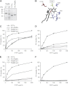
Comparison of PI and PC transfer by Class I and II PITPs. A, recombinant human PITPα, human RdgBβ-sp1 (long form) and RdgBβ-sp2 (short form), and the PITP domain of Dm-RdgBα (1 μg each) were separated by SDS-PAGE and stained with Coomassie Blue to demonstrate their degree of purity. B, residues that coordinate the inositol ring of PI in the lipid binding pocket of PITPα (Lys-61, Asn-90, Thr-59, and Glu-86) are conserved in RdgBβ. The location of residue Cys-95 in PITPα is indicated. This residue is replaced by a threonine in RdgB proteins. C, PI transfer activity by PITPα and RdgB proteins using [3H]inositol-labeled microsomes as donor and liposomes as acceptors. D, PC transfer using [3H]choline-labeled microsomes as donor and liposomes as acceptors. E, PI transfer using permeabilized HL60 cells prelabeled with [3H]inositol with liposomes as acceptors. F, PC transfer using permeabilized HL60 cells prelabeled with [3H]choline with liposomes as acceptors. The results are representative of a minimum of three experiments, and two or more protein preparations were assessed. The activity of PITPα was set as 100%. Results are shown from individual experiments performed in duplicate (±range indicated by error bars).
FIGURE 3.
FIGURE 3.
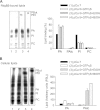
Analysis of the lipid binding specificity of RdgBβ. A, His-tagged RdgBβ and PITPα (120 μg) were incubated for 20 min with prepermeabilized HL60 cells (∼107 cells) prelabeled with [14C]acetate for 48 h. The cells were then removed by centrifugation, and RdgBβ and PITPα in the supernatant was captured using nickel beads. The lipids bound to the protein were extracted and separated by TLC. B, to monitor capture by the nickel beads, a sample of the protein (∼2 μg) was analyzed by SDS-PAGE and stained with Coomassie Blue. C, quantification of the lipid bound to PITPα (n = 5) and RdgBβ (n = 6) expressed as a percentage of total lipid binding (PC + PI + PA) to each PITP. Error bars are S.E. D, the lipid bound adjusted for the amount of recombinant protein recovered. PI bound by PITPα was set at 100%. PITPα, n = 5; RdgBβ, n = 3. Error bars are S.E. Ctrl, control.
FIGURE 4.
FIGURE 4.
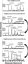
Mass spectrometric analysis of lipids associated with RdgBβ before and after exposure to HL60 cells. A, lipids extracted directly from recombinant RdgBβ expressed in E. coli. B, RdgBβ exposed to HL60 cells in the presence of bacterial phospholipase D. C, HL60 cells after exposure to RdgBβ and bacterial PLD (0.2 unit). D, RdgBβ exposed to HL60 cells in the presence of GTPγS and pCa 5 to activate endogenous PLD. E, HL60 cells after exposure to RdgBβ, GTPγS, and pCa 5.
FIGURE 5.
FIGURE 5.
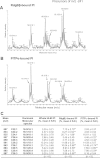
RdgBβ binds cellular PA species selectively. A, HL60 cells (5 × 10⁷ cells) were permeabilized with streptolysin O and incubated with His-tagged RdgBβ (600 μg) for 20 min at 37 °C. The cells were removed by centrifugation, and the RdgBβ present in the supernatant was recaptured with nickel beads. The lipids were extracted from the protein and analyzed by mass spectrometry. A representative electrospray ionization-MS/MS precursor scan of m/z −153 fragment in negative ionization showing the PA composition of RdgBβ-bound lipid and whole HL60 cell lipid extract is shown. B, comparison of the percent distribution of the whole cell PA molecular species with RdgBβ-bound PA (mean ± S.E. (n = 6)). RdgBβ-bound PA was also determined after HL60 cells were stimulated with GTPγS and pCa 5 to activate endogenous PLD (mean ± S.E. (n = 6)). Species identified represent the dominant species from MS/MS fragmentation analysis. A significant difference between whole cell and RdgBβ-bound PA for each molecular species is indicated by either * (p < 0.05) or ** (p < 0.005).
FIGURE 6.
FIGURE 6.
RdgBβ and PITPα prefer different molecular species of PI. HL60 cells (5 × 10⁷ cells) were permeabilized with streptolysin O, washed, and incubated with His-tagged RdgBβ or PITPα (600 μg) for 20 min at 37 °C. The cells were removed by centrifugation, and the proteins in the supernatant were captured with nickel beads. The lipids were extracted from the protein and analyzed by mass spectrometry. A representative electrospray ionization-MS/MS precursor scan of m/z −241 precursor in negative ionization showing the PI molecular species present in RdgBβ (A) and PITPα (B) is shown. C, comparison of the fractional representation of the whole cell PI molecular species with RdgBβ-bound and PITPα-bound PI. Species identified represent the dominant species from MS/MS fragmentation analysis. A significant difference between whole cell and PITP-bound PI for each molecular species is indicated by either * (p < 0.05) or ** (p < 0.005).
FIGURE 7.
FIGURE 7.
RdgBβ but not PITPα binds pyrene-labeled PA. Quenched donor vesicles consisting of a pyrene-labeled phospholipid/sn1-palmitoyl-sn2-oleylphosphatidylcholine/N-trinitrophenyl PE (2:88:10, mol/mol) were titrated with PITPα or RdgBβ. The increase in fluorescence intensity of the different pyrenyl lipid as a function of PITPα or RdgBβ added is shown in A and B, respectively. In C and D, the data are replotted to show the relative binding of PI (C) and PA (D) by PITPα versus RdgBβ. A representative experiment of three is shown.
FIGURE 8.
FIGURE 8.
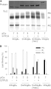
Increased PA binding by RdgBβ after stimulation of HL60 cells with GTPγS. [14C]Acetate-labeled HL60 cells were incubated with RdgBβ or PITPα in the presence of GTPγS (100 μm) or PMA (100 nm) and calcium-buffered at pCa 5 (10 μm) or pCa 7 (100 nm) together with the permeabilizing agent streptolysin O. After 20 min, the cells were removed by centrifugation, and the PITPs were captured from the supernatant using nickel beads. A, the lipids were extracted and separated by TLC, and the plate was imaged using a phosphorimaging screen. A sample of the captured proteins was also analyzed by SDS-PAGE. B, lipid binding was quantitated from two independent experiments. Error bars are S.D. (n = 2).
FIGURE 9.
FIGURE 9.
RdgBβ binds phosphatidylalcohols poorly compared with PA. HL60 cells prelabeled with [14C]acetate were incubated with RdgBβ in the presence of pCa 7 (100 nm) (control), GTPγS (100 μm) and pCa 5 (10 μm), and ethanol (2%) or butanol (BtOH) (0.5%) as indicated in the presence of streptolysin O for 20 min. A, TLC of radiolabeled lipids bound to RdgBβ and their quantitation. B, TLC of the cellular lipids and their quantitation. PBut, phosphatidylbutanol; PSL, photostimulated luminescence; PAlc, phosphatidylalcohol. The results are quantitated from four independent experiments. Error bars are S.E. (n = 4).
FIGURE 10.
FIGURE 10.
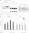
Increased PA binding to RdgBβ is dependent on endogenous phospholipase D activation and is mimicked by exogenous bacterial phospholipase D. [14C]Acetate-labeled HL60 cells were incubated with RdgBβ (120 μg) in the presence of GTPγS (100 μm), FIPI (750 nm), or U73122 (10 μm) and calcium-buffered at pCa 5 (10 μm) or pCa 7 (100 nm) as indicated together with the permeabilizing agent streptolysin O. After 20 min, the cells were removed by centrifugation and retained, and the PITPs were captured from the supernatant using nickel beads. A, the lipids were extracted and analyzed by TLC, and a sample of the captured proteins was also analyzed by SDS-PAGE. B, lipid binding to RdgBβ was quantified from three independent experiments. C, [14C]acetate-labeled HL60 cells were incubated with RdgBβ (120 μg) in the presence of streptomycin sp. PLD (0.2 unit) or GTPγS (100 μm) plus pCa 5 (10 μm) or pCa 7 (100 nm) as indicated together with the permeabilizing agent streptolysin O. After 20 min, the cells were removed by centrifugation and retained, and the PITPs were captured from the supernatant using nickel beads. Radiolabeled PA binding to RdgBβ was quantitated from three independent experiments. Error bars are S.E. (n = 3). Ctrl, control.
FIGURE 11.
FIGURE 11.
The PITP domain of Class II RdgBα proteins also binds PA. [14C]Acetate-labeled HL60 cells were incubated with the PITP domain of human (h) RdgBα, Dm-RdgBα, Dm-RdgBα(T95C), or human RdgBβ (120 μg) in the presence of GTPγS (100 μm) and calcium-buffered at pCa 5 (10 μm) or pCa 7 (100 nm) as indicated together with the permeabilizing agent streptolysin O. After 20 min, the cells were removed by centrifugation and retained, and the PITPs were captured from the supernatant using nickel beads. A, the radiolabeled lipids were extracted, separated by TLC, and imaged using a phosphorimaging screen. A sample of the captured proteins was also analyzed by SDS-PAGE. B, lipid binding was quantitated using AIDA software. The data are means of three independent experiments ±S.E. (error bars).
FIGURE 12.
FIGURE 12.
Comparison of the PI and PC transfer activity of PITP domains of RdgBα and PITPα. PI and PC transfer activity of PITPα and the PITP domains of Dm-RdgBα wild type and mutant proteins was measured using appropriately radiolabeled microsomes as donor and liposomes as acceptors. The results are representative of four independent experiments, and two or more protein preparations were assessed. The activity of PITPα was set as 100%. Error bars are S.E. hRdgBα, human RdgBα.
FIGURE 13.
FIGURE 13.
RdgBβ binds PA derived from the phospholipase D signaling pathway. The molecular species of PA bound by RdgBβ is similar to that present in PC. Other evidence to support this is the ability of the PLD inhibitor FIPI to inhibit GTPγS-stimulated PA binding to RdgBβ.

Similar articles

Cited by

References

    1. Cockcroft S., Garner K. (2011) Function of the phosphatidylinositol transfer protein gene family: is phosphatidylinositol transfer the mechanism of action? Crit. Rev. Biochem. Mol. Biol. 46, 89–117 - PubMed
    1. Allen-Baume V., Ségui B., Cockcroft S. (2002) Current thoughts on the phosphatidylinositol transfer protein family. FEBS Lett. 531, 74–80 - PubMed
    1. Yoder M. D., Thomas L. M., Tremblay J. M., Oliver R. L., Yarbrough L. R., Helmkamp G. M., Jr. (2001) Structure of a multifunctional protein. Mammalian phosphatidylinositol transfer protein complexed with phosphatidylcholine. J. Biol. Chem. 276, 9246–9252 - PubMed
    1. Tilley S. J., Skippen A., Murray-Rust J., Swigart P. M., Stewart A., Morgan C. P., Cockcroft S., McDonald N. Q. (2004) Structure-function analysis of human [corrected] phosphatidylinositol transfer protein α bound to phosphatidylinositol. Structure 12, 317–326 - PubMed
    1. Vordtriede P. B., Doan C. N., Tremblay J. M., Helmkamp G. M., Jr., Yoder M. D. (2005) Structure of PITPβ in complex with phosphatidylcholine: comparison of structure and lipid transfer to other PITP isoforms. Biochemistry 44, 14760–14771 - PubMed

Publication types