. 2012 Oct 9;109(41):E2737-46.
doi: 10.1073/pnas.1110067109. Epub 2012 Jul 20.

Damaged DNA induced UV-damaged DNA-binding protein (UV-DDB) dimerization and its roles in chromatinized DNA repair

Affiliations

Damaged DNA induced UV-damaged DNA-binding protein (UV-DDB) dimerization and its roles in chromatinized DNA repair

Joanne I Yeh et al. Proc Natl Acad Sci U S A. .

Abstract

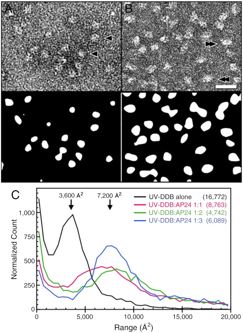
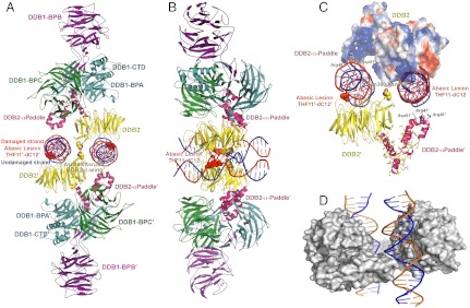
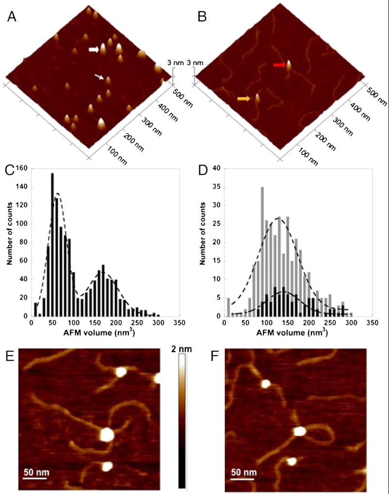
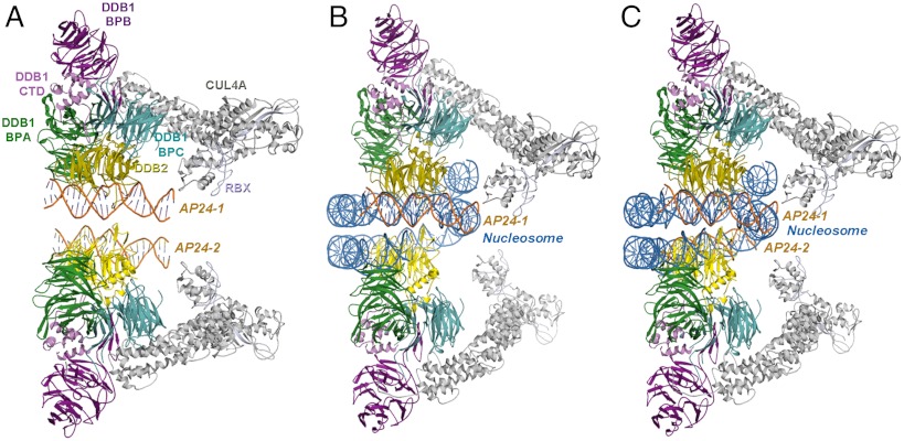
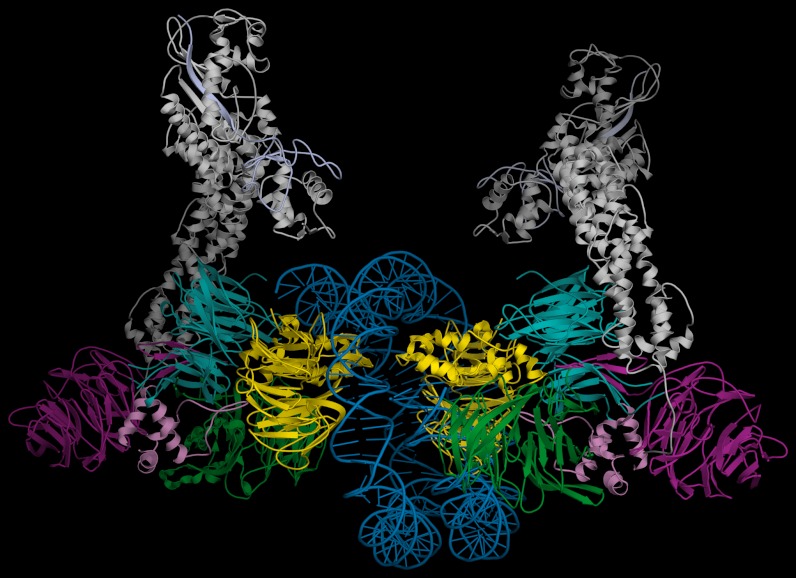
UV light-induced photoproducts are recognized and removed by the nucleotide-excision repair (NER) pathway. In humans, the UV-damaged DNA-binding protein (UV-DDB) is part of a ubiquitin E3 ligase complex (DDB1-CUL4A(DDB2)) that initiates NER by recognizing damaged chromatin with concomitant ubiquitination of core histones at the lesion. We report the X-ray crystal structure of the human UV-DDB in a complex with damaged DNA and show that the N-terminal domain of DDB2 makes critical contacts with two molecules of DNA, driving N-terminal-domain folding and promoting UV-DDB dimerization. The functional significance of the dimeric UV-DDB [(DDB1-DDB2)(2)], in a complex with damaged DNA, is validated by electron microscopy, atomic force microscopy, solution biophysical, and functional analyses. We propose that the binding of UV-damaged DNA results in conformational changes in the N-terminal domain of DDB2, inducing helical folding in the context of the bound DNA and inducing dimerization as a function of nucleotide binding. The temporal and spatial interplay between domain ordering and dimerization provides an elegant molecular rationale for the unprecedented binding affinities and selectivities exhibited by UV-DDB for UV-damaged DNA. Modeling the DDB1-CUL4A(DDB2) complex according to the dimeric UV-DDB-AP24 architecture results in a mechanistically consistent alignment of the E3 ligase bound to a nucleosome harboring damaged DNA. Our findings provide unique structural and conformational insights into the molecular architecture of the DDB1-CUL4A(DDB2) E3 ligase, with significant implications for the regulation and overall organization of the proteins responsible for initiation of NER in the context of chromatin and for the consequent maintenance of genomic integrity.


Conflict of interest statement

The authors declare no conflict of interest.

Figures

Fig. 1.
Fig. 1.
Visualization and size estimation of UV-DDB particles by negative stain electron microscopy. Representative areas are shown in A without DNA and in B with AP24 oligodeoxynucleotide at a ratio of 1∶3. (Top) Images from electron micrographs and (Bottom) after global and local filtering and thresholding to yield countable particle areas. (C) Histograms collected from micrographs of particle areas for different ratios of UV-DDB to AP24 oligodeoxynucleotide, as indicated, and normalized by particle count (in parentheses). The peak at approximately 36 nm2 evident in the absence of DNA corresponds to a circle of diameter approximately 7 nm that is consistent with a monomer of the UV-DDB1-DDB2 complex. Increasing concentrations of AP24 oligodeoxynucleotide causes the peak shifts to approximately 72 nm2 consistent with a population of dimers. Examples of monomer-sized areas are indicated with arrowheads in A and dimers with double-arrowheads in B.
Fig. 2.
Fig. 2.
Structure of the dimeric human UV-DDB in a complex with damaged DNA. (A) The dimeric UV-DDB subunit organization, shown in ribbon depiction, with each domain colored and labeled accordingly: yellow, DDB2 β-propeller; red, DDB2 N-terminal-α paddle; blue, DDB1 BPA; green, DDB1 BPC; and purple, DDB1 BPB. The 24-bp oligodeoxynucleotide (AP24) contains an abasic lesion site (THF11), with the phosphor-deoxyribose backbone of the damaged strand colored in red and the undamaged strand colored in blue. Each DDB2 subunit is bound to an AP24 oligonucleotide, with DDB2 residues Asn360/Asn360’ straddling the twofold symmetry axis, forming H bonds across the dimer interface. The surface of the Asn360/Asn360’ pair (colored using standard atom convention) is located in a loop spanning two antiparallel β-strands (β-wing). The abasic lesion site in AP24 is marked by the surface mesh drawn around nucleotides THF11/dC12 in their flipped, extra-helical configuration. The β-wing is sandwiched between the two AP24 oligodeoxynucleotides, astride of the twofold axis of rotation relating the monomer subunits in the dimeric DDB2. Residues on the leading β-strand and loop form contacts with the undamaged DNA strand whereas residues on the loop and the retreating β-strand form contacts with the neighboring undamaged DNA strand. Both sets of contacts are predominantly electrostatic in nature, thus largely sequence independent. (B) Same as A but rotated 90 degrees and tilted slightly to show both DNA molecules. (C) Electrostatic potential surfaces of the DDB2 N-terminal domain complement the charge characteristics of the DDB1 BPC domain and the DNA phosphor-deoxyribose interfaces, resulting in favorable electrostatic neutralization. Contacts between residues on the β-wing region form contacts with the DNA bound at its immediate active site and with the neighbouring DNA molecule bound to the second monomer of DDB2 in the dimer. Extensive interactions between residues on the N-terminal-helical domain (α-paddle) and the neighboring DNA molecule augment the intermolecular associations, contributing to the high affinity of damaged DNA binding. (D) The skewed positioning of the DNA binding surface can now be understood in terms of the DDB2 dimer interface, located adjacent to the DNA binding site, at a loop bridging blades 6 and 7 of the β-propeller (β-wing) of DDB2. To accommodate the steric constraints imposed through dimerization along with DNA binding, the two adjacent sites are positioned diametrically across one face of the molecular surface of DDB2, readily seen in the dimeric DDB2-DNA (AP24) complex.
Fig. 3.
Fig. 3.
AFM imaging shows that damaged DNA binding promotes the dimerization of the DDB1-DDB2 heterodimer. (A) A representative surface plot of UV-DDB (50 nM) in the absence of DNA. The thin and wide white arrows point to molecules consistent with the size of the UV-DDB monomer (DDB1-DDB2 heterodimer) and trimer of UV-DDB, respectively. (B) Representative surface plot of UV-DDB (50 nM) in the presence of UV-irradiated 517 bp PCR fragments (25 nM). The yellow and red arrows point to dimeric UV-DDB [(DDB1-DDB2)2] binding to one and two molecules of duplex DNA, respectively. (C) AFM volume analysis of free UV-DDB (n = 1,160). (D) AFM volume analysis of UV-DDB on one strand (gray bars, n = 339) and two strands (black bars, n = 79) of duplex DNA. The images in A and B are at 500 nm × 500 nm and 3 nm in height. (Bottom) The dashed lines (C, free in solution, and D, bound to DNA) represent Gaussian fits to the data. Field view images of UV-DDB binding to separate DNA molecules (E) or two different regions of the same DNA molecule (F). The images are at 300 nm × 300 nm and 2 nm in height.
Fig. 4.
Fig. 4.
Model of DDB1-CUL4ADDB2 ubiquitin ligase complexed to a nucleosome. (A) Modeling of the complex with CUL4A-Rbx (gray, light blue) onto the dimeric UV-DDB2 (domains colored as in Fig. 2); the region defined by two adjacent AP24 oligodeoxynucleotides (AP24-1 & AP24-2, in orange) used for the docking of a nucleosome; (B) Docking of the nucleosome in the dimeric UV-DDB, showing the fit of one AP24-1 (in orange) relative to the nucleosome (in blue); (C) fit of the nucleosome onto both oligomers showing that the distance between the two oligonucleotides can readily accommodate the nucleosome molecule (second DNA molecule, AP24-2, shown in orange) with minor adjustments of the second DDB2 component, as needed. The dimeric scaffold accommodates the numerous proteins that transiently assemble and disassemble on the DDB1-CUL4ADDB2 ubiquitin ligase complex at the vicinity of the lesion site in the subsequent DNA repair process. The dimeric architecture also spatially aligns the various molecular subunits in the reactions monoubiquitinylating histones and polyubiquitinylating substrate receptors (i.e., DDB2) for proteasomal degradation and verified by docking the E2 ubiquitin transferase enzyme onto the DDB1-CUL4ADDB2 ubiquitin ligase complex, resulting in E2 bridging distances to histones. In this figure, the histone and E2 proteins are omitted for clarity.
Fig. P1.
Fig. P1.
Composite model of a dimeric DDB1-CUL4ADDB2 ubiquitin ligase-nucleosome complex. A model of a dimeric DDB1-CUL4ADDB2 in a complex with a nucleosome core particle, generated according to the relative subunit organization found in the dimeric UV-DDB-AP24 crystal structure. The intermolecular orientation of the CUL4A-Rbx (in gray) subunit resulted from superimposing two copies of the respective BPA and BPC domains of the DDB1 subunit in the DDB1-CUL4A-Rbx structure (accession number 2HYE) onto the corresponding DDB1 domains (BPA in blue, BPC in green, BPB in purple) in the dimeric UV-DDB crystal structure. The nucleosome core particle (PDB ID code 1AOI) was aligned by superpositioning the respective sugar-phosphate backbones of the nucleosomal DNA (in blue) onto the backbones of two adjacent AP24 oligodeoxynucleotides (not shown). Remarkably, the available molecular volume between two AP24 DNA molecules accommodates the spatial requirements of the nucleosome particle, with minor adjustments of the two DDB2 subunits (in yellow). For visual clarity, the histones of the nucleosome core particle and the AP24 subunits are not depicted in this figure. For a view showing the superpositioning of the nucleosome onto the two AP24 oligodeoxynucleotide molecules of the UV-DDB complex, refer to Fig. 4 of the complete on-line publication.

References

    1. Friedberg EC, et al. DNA Repair and Mutagenesis. 2nd Ed. Washington, DC: ASM Press; 2005. pp. 1–1164.
    1. Sugasawa K, et al. Xeroderma pigmentosum group C protein complex is the initiator of global genome nucleotide excision repair. Mol Cell. 1998;2:223–232. - PubMed
    1. Wittschieben BO, Wood RD. DDB complexities. DNA Repair (Amst) 2003;2:1065–1069. - PubMed
    1. Moser J, et al. The UV-damaged DNA binding protein mediates efficient targeting of the nucleotide excision repair complex to UV-induced photo lesions. DNA Repair (Amst) 2005;4:571–582. - PubMed
    1. Wakasugi M, et al. DDB accumulates at DNA damage sites immediately after UV irradiation and directly stimulates nucleotide excision repair. J Biol Chem. 2002;277:1637–1640. - PubMed

Publication types

MeSH terms