. 2012 Dec;11(6):1121-4.
doi: 10.1111/j.1474-9726.2012.00866.x. Epub 2012 Aug 27.

Chromosome cohesion decreases in human eggs with advanced maternal age

Affiliations

Chromosome cohesion decreases in human eggs with advanced maternal age

Francesca E Duncan et al. Aging Cell. 2012 Dec.

Abstract

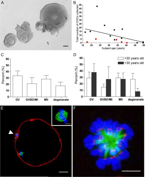
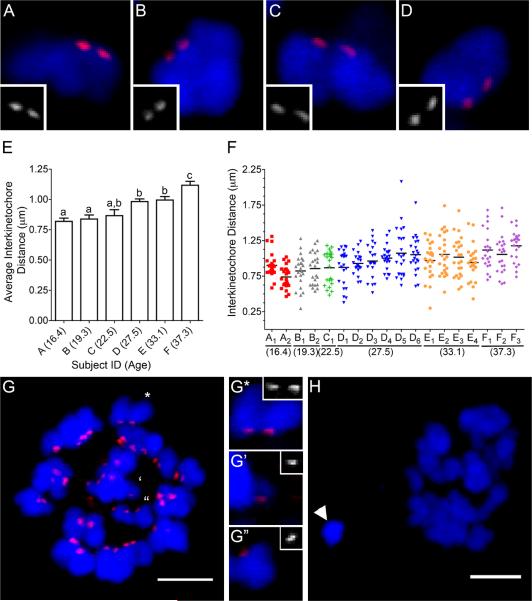
Aneuploidy in human eggs increases with maternal age and can result in infertility, miscarriages, and birth defects. The molecular mechanisms leading to aneuploidy, however, are largely unknown especially in the human where eggs are exceedingly rare and precious. We obtained human eggs from subjects ranging from 16.4 to 49.7 years old following in vitro maturation of oocyte-cumulus complexes isolated directly from surgically removed ovarian tissue. A subset of these eggs was used to investigate how age-associated aneuploidy occurs in the human. The inter-kinetochore distance between sister chromatids increased significantly with maternal age, indicating weakened cohesion. Moreover, we observed unpaired sister chromatids from females of advanced age. We conclude that loss of cohesion with increasing maternal age likely contributes to the well-documented increased incidence of aneuploidy.


Figures

Figure 1
Figure 1
Human MII eggs can be obtained following IVM of OCCs isolated directly from surgically-removed ovarian tissue. (A) Morphology of OCCs released from small antral follicles during ovarian tissue processing for cryopreservation. Scale bar = 100 μm. (B) The effects of age and previous cancer therapy (red squares) on the number of OCCs collected per subject. (C–D) The percent of cells in each stage of meiotic progression following IVM for 36–42 hours (GV, germinal vesicle-intact; GVBD/MI, germinal vesicle breakdown/metaphase of meiosis l; MII, metaphase of meiosis II) reported as an (C) overall total or (D) separated according to age (> or < 30 years old). There were no statistical differences between the groups. (E) The cytoskeleton in a human MII egg following IVM. (F-actin = red; α-tubulin = green, DNA = blue). The polar body is marked by an arrowhead and the inset highlights the bipolar metaphase II spindle. Scale bar = 25 μm. (F) The human meiotic spindle following monastrol treatment (α-tubulin = green, DNA = blue, kinetochores = red). Scale bar = 5 μm.
Figure 2
Figure 2
Inter-kinetochore distances increase significantly and chromosome segregation errors occur more frequently in human eggs with advanced age. Representative sister chromatids from (A–B) Subject D (27.5 years old) has on average smaller inter-kinetochore distances compared to those from (C–D) Subject F (37.3 years old). (E) The average inter-kinetochore distances of eggs from individual subjects are plotted with increasing maternal age. The uppercase letters correspond to the subject ID, and the subject age in years is indicated in parentheses. Different lowercase letters denote significant differences. (F) All the measured inter-kinetochore distances per individual egg are plotted with increasing maternal age. The uppercase letters, consistent with the labeling in (E), represent the subject ID and the subscript number indicates an individual egg. Each color represents inter-kinetochore measurements from the same subject. The mean inter-kinetochore distance for each egg is indicated by a black line. Eggs B1, E3, E4, F1, and F2 were determined to have chromosome segregation errors. (G) An egg from Subject F (F2) with a pair of separated sister chromatids. The marked chromosomes are further magnified to show a representative image of an intact sister chromatid pair (G*) and the pair of separated sister chromatids (G', G”; separated by four 0.2 μm-thick optical sections. (H) Another egg from Subject F (F1) had a chromosome pair (arrowhead) that was separated from the egg DNA (**) in both the XY-plane and Z-plane (0.8 μm). Kinetochores = red and DNA = blue. The scale bar = 5 μm.

References

    1. Chiang T, Duncan FE, Schindler K, Schultz RM, Lampson MA. Evidence that weakened centromere cohesion is a leading cause of age-related aneuploidy in oocytes. Curr Biol. 2010;20:1522–1528. - PMC - PubMed
    1. Chiang T, Schultz RM, Lampson MA. Age-dependent susceptibility of chromosome cohesion to premature separase activation in mouse oocytes. Biol Reprod. 2011;85:1279–1283. - PMC - PubMed
    1. Chiang T, Schultz RM, Lampson MA. Meiotic origins of maternal age-related aneuploidy. Biol Reprod. 2012;86:1–7. - PMC - PubMed
    1. Duncan FE, Chiang T, Schultz RM, Lampson MA. Evidence that a defective spindle assembly checkpoint is not the primary cause of maternal age-associated aneuploidy in mouse eggs. Biol Reprod. 2009;81:768–776. - PMC - PubMed
    1. Eichenlaub-Ritter U, Staubach N, Trapphoff T. Chromosomal and cytoplasmic context determines predisposition to maternal age-related aneuploidy: overview and update on MCAK in mammalian oocytes. Biochem Soc Trans. 2010;38:1681–1686. - PubMed

Publication types