Review
. 2012 Jul 23:12:304.
doi: 10.1186/1471-2407-12-304.

Myeloid malignancies: mutations, models and management

Affiliations
Review

Myeloid malignancies: mutations, models and management

Anne Murati et al. BMC Cancer. .

Abstract

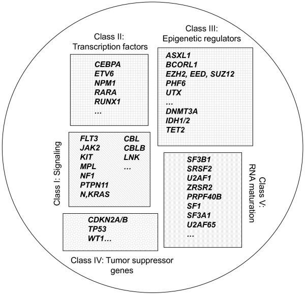
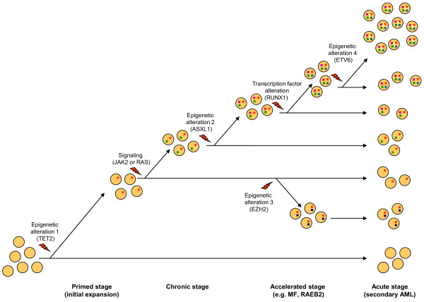
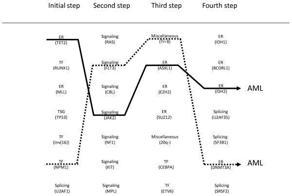
Myeloid malignant diseases comprise chronic (including myelodysplastic syndromes, myeloproliferative neoplasms and chronic myelomonocytic leukemia) and acute (acute myeloid leukemia) stages. They are clonal diseases arising in hematopoietic stem or progenitor cells. Mutations responsible for these diseases occur in several genes whose encoded proteins belong principally to five classes: signaling pathways proteins (e.g. CBL, FLT3, JAK2, RAS), transcription factors (e.g. CEBPA, ETV6, RUNX1), epigenetic regulators (e.g. ASXL1, DNMT3A, EZH2, IDH1, IDH2, SUZ12, TET2, UTX), tumor suppressors (e.g. TP53), and components of the spliceosome (e.g. SF3B1, SRSF2). Large-scale sequencing efforts will soon lead to the establishment of a comprehensive repertoire of these mutations, allowing for a better definition and classification of myeloid malignancies, the identification of new prognostic markers and therapeutic targets, and the development of novel therapies. Given the importance of epigenetic deregulation in myeloid diseases, the use of drugs targeting epigenetic regulators appears as a most promising therapeutic approach.


Figures

Figure 1
Figure 1
Circos diagrams depict the relative frequency and associations of the major mutations in MPNs (a) and MDSs (b), respectively based on data from our work[37]on127 classic MPNs and from Damm’s study[38]on 221 MDSs. Wild-type means that no disease allele has been detected in the genes listed.
Figure 2
Figure 2
Schematic representation of five classes of leukemogenic genes. ERs (class III) can be subdivided into two subclasses (DNA methylation-associated and histone-associated).
Figure 3
Figure 3
Schematic representation of epigenetic regulation of a leukemogenic locus (LL) framed by histone H3. (a) Histone acetyltransferases (HAT; e.g. MYST3) and histone methyltransferases (HMT; e.g. MLL) can activate the locus. (b) Reciprocally, the locus is repressed by polycomb complex PRC2 (which comprised EED, EZH2 and SUZ12 proteins). ASXL1 would direct PRC2 to the locus. Loss-of-function mutations in PRC2 components or in ASXL1 remove PRC2 repression. DNMT3A is involved in the formation of 5-methylcytosines (5mC) from cytosines and interacts with HMTs as well as with PRC2 components. TET2 mediates hydroxylation of 5mC to 5hmC. To function, TET2 requires α-ketoglutarate (αKG), which is provided by IDH1/2 proteins. Aberrant methylation patterns are caused by mutation in TET2 or in IDH1/2, which produces 2-hydroxyglutarate instead of αKG.
Figure 4
Figure 4
Schematic representation of pathways leading to acute myeloid leukemia (AML) from hematopoietic stem cell (HSC) or progenitors (PG). Gene fusions and NPM1 mutation are major events in the induction of primary AMLs with favorable and intermediate cytogenetic risk (they correspond respectively to mutation groups A and B of Ley et al [24], and to mutation groups 2 + 3 and 1 of Shen et al. [101]. Secondary AML following MPN or MDS (see Figure 1) could occur after a series of gene mutations in transcription factors and epigenetic regulators combined with a mutation in a signaling pathway (see Figure 3), after TP53 mutation and a series of mutations and karyotype alterations due to genetic instability, or after additional mutations in BCR-ABL chronic myeloid leukemia.
Figure 5
Figure 5
Schematic representation of a case of malignant myeloid disease evolving in four stages along one pathway. Clones with different gene mutations (color squares in cells) represent various ratios of the oligoclonal leukemia. The order and nature of the mutations (or genome alterations) is given as an example and may differ from one case to another. However, in contrast to JAK2V617F, which has a mild effect on hematopoietic stem cell (HSC) [16], TET2 mutation has the property to initiate the amplification of HSC and to pave the way to secondary mutations [77]. Mutations in signaling molecules, which have a major impact on the disease phenotype, will vary with the type of chronic stage, for example it could affect JAK2 in case of MPN, RAS in case of MP-CMML and be absent in case of MDS. MF: myelofibrosis, RAEB: refractory anemia with excess of blasts, AML: acute myeloid leukemia.
Figure 6
Figure 6
“Slot machine” model of leukemogenesis. Alterations in signaling molecules, transcription factors (TFs), epigenetic regulators (ERs), tumor suppressors (TSG), spliceosome components and various genome abnormalities (examples are given) fall into (at least) four “reels” (steps) that combine to induce a malignant myeloid disease. Acute myeloid leukemia (AML) results from one of the allowed combinations of four (at least) cooperating alterations. At chronic stages, the steps are variably combined, some may be absent (e.g. signaling), some may be specific (e.g. SF3B1 splicing mutations in RARS). Each step can be achieved by alterations in one of several genes. The initial step leads to expansion of a founding clone. Two examples of draw (plain and dotted lines) leading to AML are shown.

Similar articles

Cited by

References

    1. Chen J, Odenike O, Rowley JD. Leukaemogenesis: more than mutant genes. Nat Rev Cancer. 2010;10:23–36. - PMC - PubMed
    1. Baxter EJ, Scott LM, Campbell PJ, East C, Fourouclas N, Swanton S, Vassiliou GS, Bench AJ, Boyd EM, Curtin N, Scott MA, Erber WN, Green AR. Acquired mutation of the tyrosine kinase JAK2 in human myeloproliferative disorders. Lancet. 2005;365:1054–1061. - PubMed
    1. James C, Ugo V, Le Couedic JP, Staerk J, Delhommeau F, Lacout C, Garcon L, Raslova H, Berger R, Bennaceur-Griscelli A, Villeval JL, Constantinescu SN, Casadevall N, Vainchenker W. A unique clonal JAK2 mutation leading to constitutive signalling causes polycythaemia vera. Nature. 2005;434:1144–1148. - PubMed
    1. Kralovics R, Passamonti F, Buser AS, Teo SS, Tiedt R, Passweg JR, Tichelli A, Cazzola M, Skoda RC. A gain-of-function mutation of JAK2 in myeloproliferative disorders. N Engl J Med. 2005;352:1779–1790. - PubMed
    1. Levine RL, Wadleigh M, Cools J, Ebert BL, Wernig G, Huntly BJ, Boggon TJ, Wlodarska I, Clark JJ, Moore S, Adelsperger J, Koo S, Lee JC, Gabriel S, Mercher T, D'Andrea A, Frohling S, Dohner K, Marynen P, Vandenberghe P, Mesa RA, Tefferi A, Griffin JD, Eck MJ, Sellers WR, Meyerson M, Golub TR, Lee SJ, Gilliland DG. Activating mutation in the tyrosine kinase JAK2 in polycythemia vera, essential thrombocythemia, and myeloid metaplasia with myelofibrosis. Cancer Cell. 2005;7:387–397. - PubMed

Publication types

MeSH terms