

Genome-wide analysis of the sox family in the calcareous sponge Sycon ciliatum: multiple genes with unique expression patterns
- PMID: 22824100
- PMCID: PMC3495037
- DOI: 10.1186/2041-9139-3-14
Genome-wide analysis of the sox family in the calcareous sponge Sycon ciliatum: multiple genes with unique expression patterns
Abstract
Background: Sox genes are HMG-domain containing transcription factors with important roles in developmental processes in animals; many of them appear to have conserved functions among eumetazoans. Demosponges have fewer Sox genes than eumetazoans, but their roles remain unclear. The aim of this study is to gain insight into the early evolutionary history of the Sox gene family by identification and expression analysis of Sox genes in the calcareous sponge Sycon ciliatum.
Methods: Calcaronean Sox related sequences were retrieved by searching recently generated genomic and transcriptome sequence resources and analyzed using variety of phylogenetic methods and identification of conserved motifs. Expression was studied by whole mount in situ hybridization.
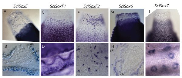
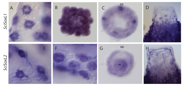
Results: We have identified seven Sox genes and four Sox-related genes in the complete genome of Sycon ciliatum. Phylogenetic and conserved motif analyses showed that five of Sycon Sox genes represent groups B, C, E, and F present in cnidarians and bilaterians. Two additional genes are classified as Sox genes but cannot be assigned to specific subfamilies, and four genes are more similar to Sox genes than to other HMG-containing genes. Thus, the repertoire of Sox genes is larger in this representative of calcareous sponges than in the demosponge Amphimedon queenslandica. It remains unclear whether this is due to the expansion of the gene family in Sycon or a secondary reduction in the Amphimedon genome. In situ hybridization of Sycon Sox genes revealed a variety of expression patterns during embryogenesis and in specific cell types of adult sponges.
Conclusions: In this study, we describe a large family of Sox genes in Sycon ciliatum with dynamic expression patterns, indicating that Sox genes are regulators in development and cell type determination in sponges, as observed in higher animals. The revealed differences between demosponge and calcisponge Sox genes repertoire highlight the need to utilize models representing different sponge lineages to describe sponge development, a prerequisite for deciphering evolution of metazoan developmental mechanisms.
Figures


Similar articles
-
Calcisponges have a ParaHox gene and dynamic expression of dispersed NK homeobox genes.Nature. 2014 Oct 30;514(7524):620-3. doi: 10.1038/nature13881. Nature. 2014. PMID: 25355364
-
Conservation and divergence of bHLH genes in the calcisponge Sycon ciliatum.Evodevo. 2016 Oct 14;7:23. doi: 10.1186/s13227-016-0060-8. eCollection 2016. Evodevo. 2016. PMID: 27757221 Free PMC article.
-
Surprisingly rich repertoire of Wnt genes in the demosponge Halisarca dujardini.BMC Evol Biol. 2016 Jun 10;16(1):123. doi: 10.1186/s12862-016-0700-6. BMC Evol Biol. 2016. PMID: 27287511 Free PMC article.
-
Comparative analyses of developmental transcription factor repertoires in sponges reveal unexpected complexity of the earliest animals.Mar Genomics. 2015 Dec;24 Pt 2:121-9. doi: 10.1016/j.margen.2015.07.008. Epub 2015 Aug 5. Mar Genomics. 2015. PMID: 26253310 Review.
-
From traveler to homebody: Which signaling mechanisms sponge larvae use to become adult sponges?Adv Protein Chem Struct Biol. 2019;116:421-449. doi: 10.1016/bs.apcsb.2019.02.002. Epub 2019 Mar 14. Adv Protein Chem Struct Biol. 2019. PMID: 31036299 Review.
Cited by
-
Calcisponges have a ParaHox gene and dynamic expression of dispersed NK homeobox genes.Nature. 2014 Oct 30;514(7524):620-3. doi: 10.1038/nature13881. Nature. 2014. PMID: 25355364
-
The ancestral gene repertoire of animal stem cells.Proc Natl Acad Sci U S A. 2015 Dec 22;112(51):E7093-100. doi: 10.1073/pnas.1514789112. Epub 2015 Dec 7. Proc Natl Acad Sci U S A. 2015. PMID: 26644562 Free PMC article.
-
Whole genome sequence of the deep-sea sponge Geodia barretti (Metazoa, Porifera, Demospongiae).G3 (Bethesda). 2023 Sep 30;13(10):jkad192. doi: 10.1093/g3journal/jkad192. G3 (Bethesda). 2023. PMID: 37619978 Free PMC article.
-
Embryonic expression patterns and phylogenetic analysis of panarthropod sox genes: insight into nervous system development, segmentation and gonadogenesis.BMC Evol Biol. 2018 Jun 8;18(1):88. doi: 10.1186/s12862-018-1196-z. BMC Evol Biol. 2018. PMID: 29884143 Free PMC article.
-
Expression of multiple Sox genes through embryonic development in the ctenophore Mnemiopsis leidyi is spatially restricted to zones of cell proliferation.Evodevo. 2014 Apr 24;5:15. doi: 10.1186/2041-9139-5-15. eCollection 2014. Evodevo. 2014. PMID: 24834317 Free PMC article.
References
-
- Gubbay J, Collignon J, Koopman P, Capel B, Economou A, Munsterberg A, Vivian N, Goodfellow P, Lovell-Badge R. A gene mapping to the sex-determining region of the mouse Y chromosome is a member of a novel family of embryonically expressed genes. Nature. 1990;346:245–250. doi: 10.1038/346245a0. - DOI - PubMed
LinkOut - more resources
Full Text Sources
Miscellaneous
