. 2012 Nov 13;26(17):2135-44.
doi: 10.1097/QAD.0b013e328357f5ad.

Long-acting nanoformulated antiretroviral therapy elicits potent antiretroviral and neuroprotective responses in HIV-1-infected humanized mice

Affiliations

Long-acting nanoformulated antiretroviral therapy elicits potent antiretroviral and neuroprotective responses in HIV-1-infected humanized mice

Prasanta K Dash et al. AIDS. .

Abstract

Objectives: Long-acting nanoformulated antiretroviral therapy (nanoART) with improved pharmacokinetics, biodistribution and limited systemic toxicities will likely improve drug adherence and access to viral reservoirs.

Design: Atazanavir and ritonavir crystalline nanoART were formulated in a poloxamer-188 excipient by high-pressure homogenization. These formulations were evaluated for antiretroviral and neuroprotective activities in humanized NOD/scid-IL-2Rgc (NSG) mice.

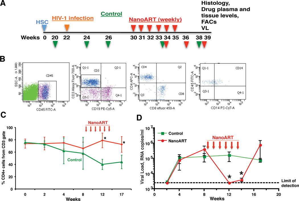
Methods: NanoART-treated NSG mice were evaluated for drug biodistribution, pharmacodynamics and toxicity. CD34 human hematopoietic stem cells were transplanted at birth in replicate NSG mice. The mice were infected with HIV-1ADA at 5 months of age. Eight weeks later, the infected animals were treated with weekly subcutaneous injections of nanoformulated ATV and RTV. Peripheral viral load, CD4 T-cell counts and lymphoid and brain histopathology and immunohistochemistry tests were performed.

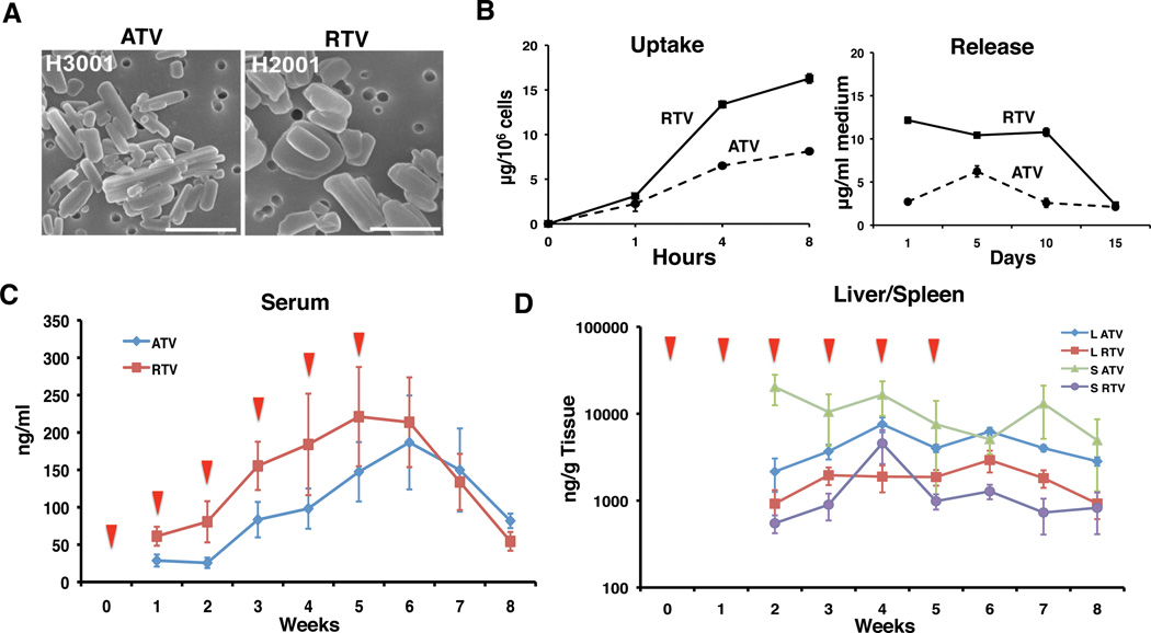
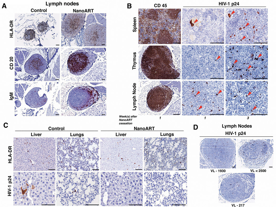
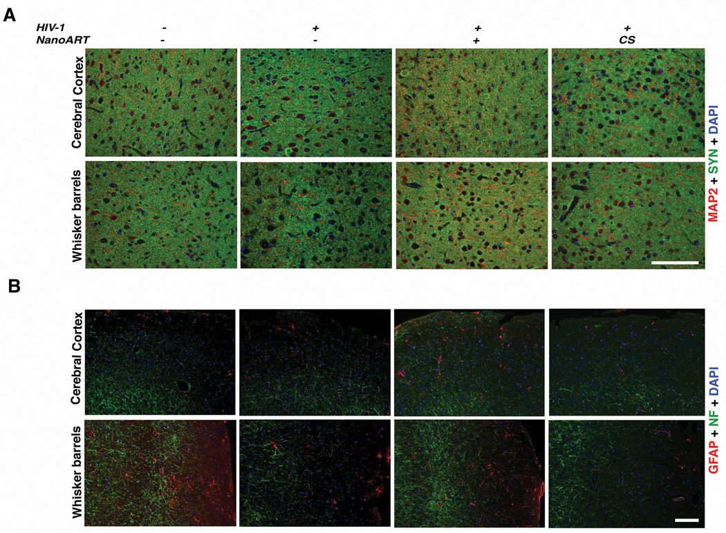
Results: NanoART treatments by once-a-week injections reduced viral loads more than 1000-fold and protected CD4 T-cell populations. This paralleled high ART levels in liver, spleen and blood that were in or around the human minimal effective dose concentration without notable toxicities. Importantly, examination of infected brain subregions showed that nanoART elicited neuroprotective responses with detectable increases in microtubule-associated protein-2, synaptophysin and neurofilament expression when compared to untreated virus-infected animals. Therapeutic interruptions produced profound viral rebounds.

Conclusion: Long-acting nanoART has translational potential with sustained and targeted efficacy and with limited systemic toxicities. Such success in drug delivery and distribution could improve drug adherence and reduce viral resistance in infected people.


Conflict of interest statement

The author reports no conflicts of interest.

Figures

Figure 1
Figure 1. NanoART characterization and tissue distribution
(A) Scanning electron micrographs (15,000×) of nanoformulations on top of a 0.2 µm polycarbonate filter membrane are shown, bar = 1 µm. (B) Time course of nanoART uptake and release of monocyte-derived macrophages. Concentration in cell lysates and media were determined by RP-HPLC. Data is expressed as mean + SEM for n = 3 determinations. (C) Measurement of ATV and RTV concentrations in the serum by UPLC-MS/MS and (D) in liver (L) and spleen (S) (log10 scale) during the study time course. Uninfected 10-week old NSG mice were injected weekly subcutaneously for 6 weeks with nanoformulations of ATV and RTV (1:1) at dosages of 250 mg/kg. A group of 5 mice were sacrificed weekly and 3 weeks after drug cessation. Drug levels were quantified in sera, liver and spleen. In sera drug was detected from week-1, and in liver and spleen, drug was detected from week 2 as shown in D. Data are expressed as medians at the 25th and 75th percentiles for n = 5. Red triangles indicate nanoART injection time.
Figure 2
Figure 2. NanoART preserves CD4+ T cells and suppresses VL
(A) Experimental scheme for human CD34+ HSC reconstitution, HIV-1 infection, nanoART administration (above time line), and viral and immune profiling (green and red triangles below time line), drug concentration and histological evaluations are shown. (B) Flow cytometric plots for human cell profiles in mouse blood. Mice were bled from facial vein once every four weeks starting the second week after HIV-1 infection and peripheral blood stained for expression of human CD45. Gated human CD45+ mononuclear cells were assessed for expression of human CD3 (T cells), CD19 (B cells) and CD14 (monocytes). CD3+ T cells were gated to assess the expression of CD4 and CD8. (C) Circulating human CD4+ T cells and (D) viral loads (VL) in peripheral blood during infection in untreated (control) animals and nanoART-treated mice at times (weeks) post-HIV-1 infection. VL, with rebound at 3 weeks of drug cessation and preserved CD4+ T cell counts in nanoART treated animals was found. Data are expressed as mean + SEM for n = 5. *Significantly different at p<0.05 compared to control.
Figure 3
Figure 3. NanoART reduces number of infected cells and restores tissue integrity
(A) Histopathology of lymph nodes in HIV-1 infected humanized mice after nanoART treatment shows reductions in follicle size with evident HLA-DR and HIV-1p24+ staining. Staining of lymph nodes with anti-CD20 and anti-IgM shows the presence of mature cells in control and nanoART animals. (B) Histopathology of lymphoid organs of hu-NSG mice one and three weeks after cessation of nanoART. ATV and RTV nanoformulations were administered starting eight weeks after viral infection. Representative sections of spleen, thymus and lymph nodes were stained for human CD45 and HIV-1 p24 antigens (brown). HIV-1 p24+ cells, readily seen (red arrows) in splenic lymphoid follicles, thymus and lymph nodes, were affected following therapy and therapeutic interruption. (C) Representative liver and lung sections were stained for human HLA-DR and HIV-1 p24 in hu-NSG mice. Untreated animals showing HIV-1p24+ cells compared to nanoART treated animals. (D) Lymph nodes of nanoART treated mice showing numbers of HIV-1p24+ cells (brown-stained). Serum VLs are shown beneath each micrograph. The scale bar in the pictures corresponds to 100 µm.
Figure 4
Figure 4. NanoART improves neuronal, synaptic and astrocyte markers damaged following HIV-1 infection
Immunofluorescence labeling of astrocytes, neurofilaments and synapses in brain regions of hu-NSG mice that were uninfected/untreated, infected/untreated, infected/nanoART-treated, and infected/nanoART-treated and suspended. Representative images captured with the Nuance multispectral imaging system are presented. Slide mounted sections (5 µm) of brain with VL ranging from very high (untreated and treatment interrupted group) to viral loads less than level of detection (group sacrificed 1 week after 2 nanoART injections) were stained for (A) dendritic MAP2, red, synaptic morphology with synaptophysin (SYN) green, and nuclei (DAPI) blue) and (B) GFAP, red, neurofilament (NF) green nuclei (DAPI) blue. Infected and nanoART treated groups have extended dendritic morphological appearance as compared to infected and treatment cessation group, where punctate structures were observed. The scale bar corresponds to 100 µm. CS is nanoART cessation.

References

    1. Volberding PA, Deeks SG. Antiretroviral therapy and management of HIV infection. Lancet. 2010;376:49–62. - PubMed
    1. Swindells S, Flexner C, Fletcher CV, Jacobson JM. The critical need for alternative antiretroviral formulations, and obstacles to their development. J Infect Dis. 2011;204:669–674. - PMC - PubMed
    1. Kadiu I, Nowacek A, McMillan J, Gendelman HE. Macrophage endocytic trafficking of antiretroviral nanoparticles. Nanomedicine (Lond) 2011;6:975–994. - PMC - PubMed
    1. Mallipeddi R, Rohan LC. Progress in antiretroviral drug delivery using nanotechnology. Int J Nanomedicine. 2010;5:533–547. - PMC - PubMed
    1. Nowacek AS, Miller RL, McMillan J, Kanmogne G, Kanmogne M, Mosley RL, et al. NanoART synthesis, characterization, uptake, release and toxicology for human monocyte-macrophage drug delivery. Nanomedicine (Lond) 2009;4:903–917. - PMC - PubMed

Publication types

MeSH terms