. 2013 Apr 4;32(14):1772-83.
doi: 10.1038/onc.2012.205. Epub 2012 Jul 23.

Tetraspanin CD151 plays a key role in skin squamous cell carcinoma

Affiliations

Tetraspanin CD151 plays a key role in skin squamous cell carcinoma

Q Li et al. Oncogene. .

Abstract

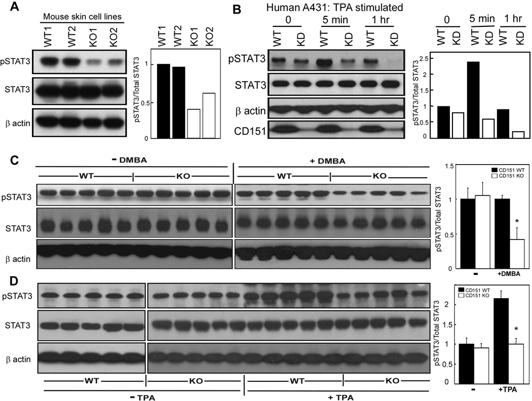
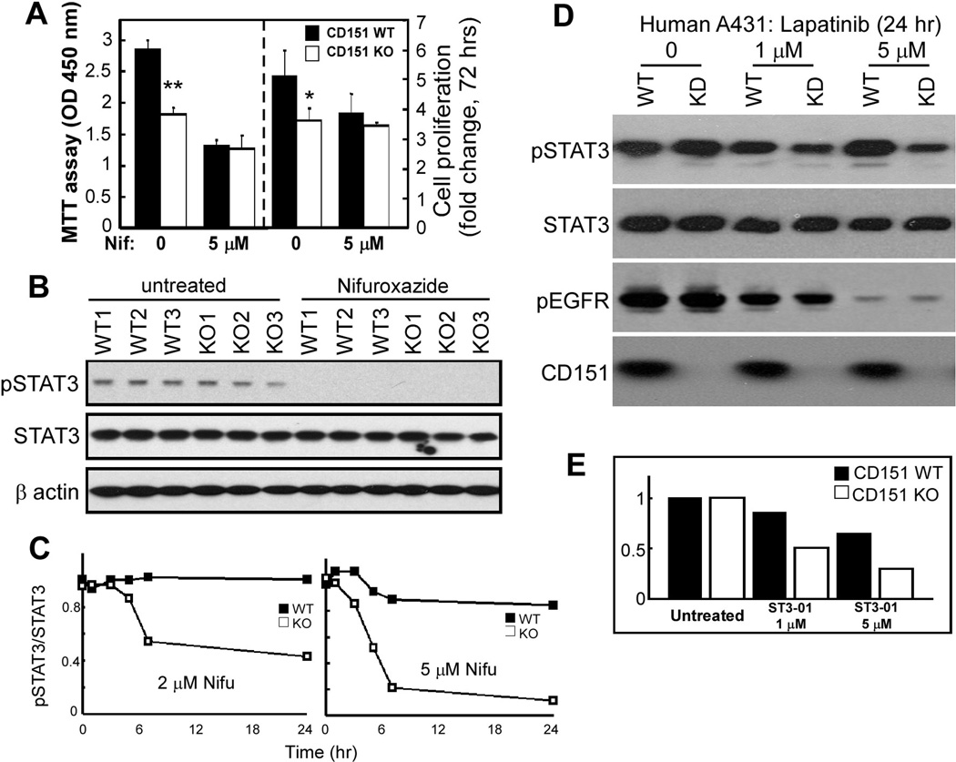
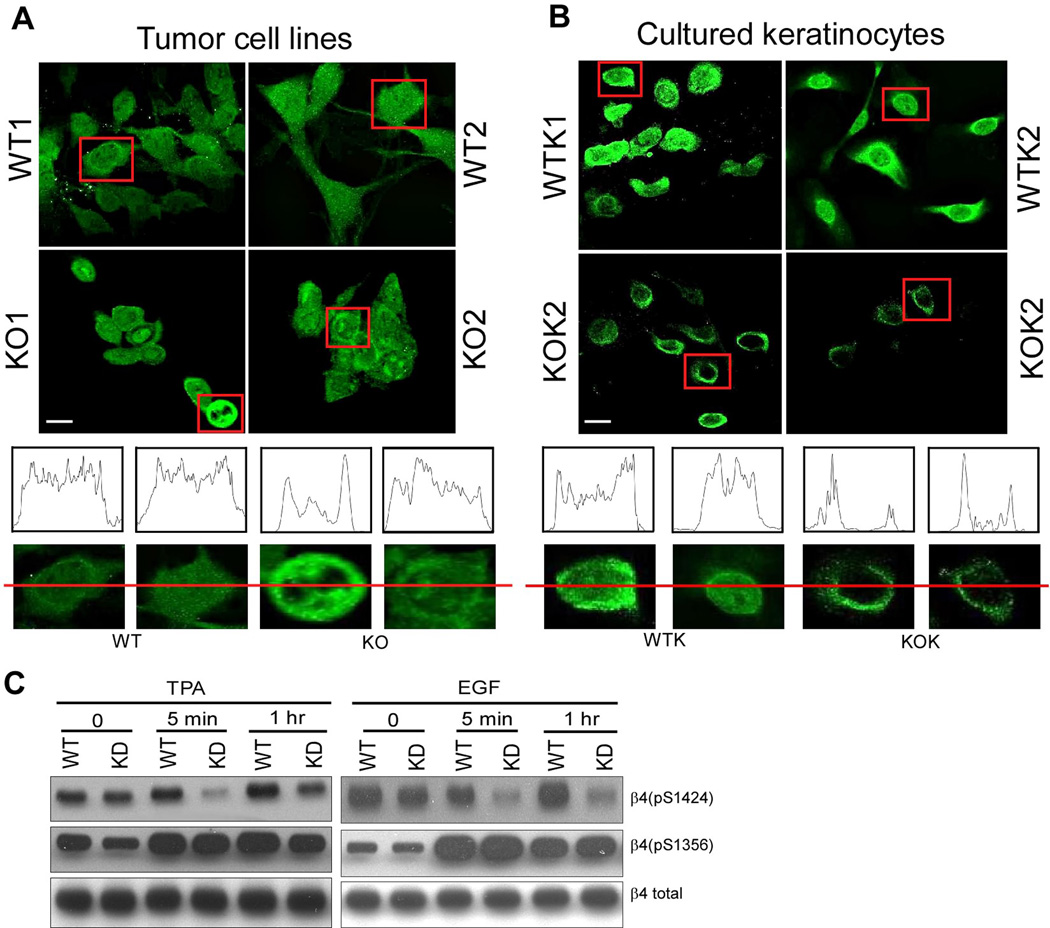
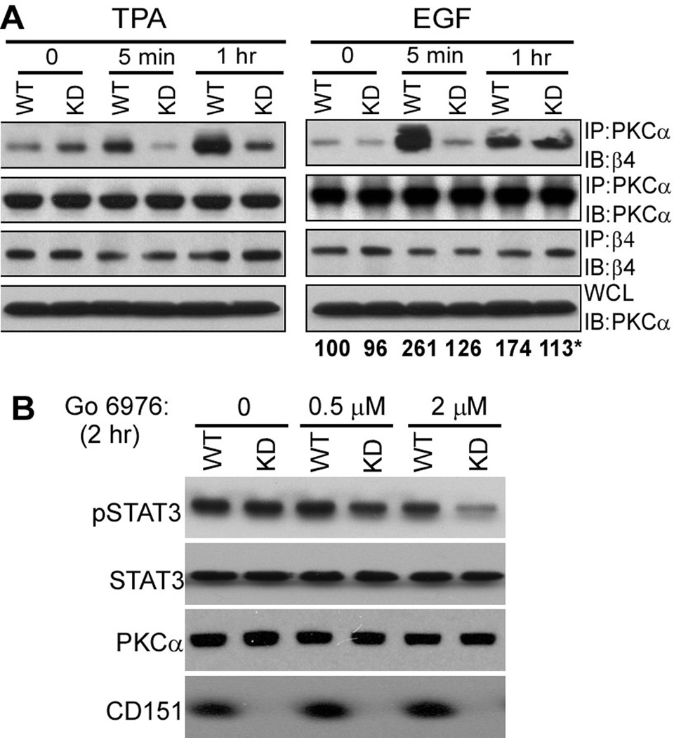
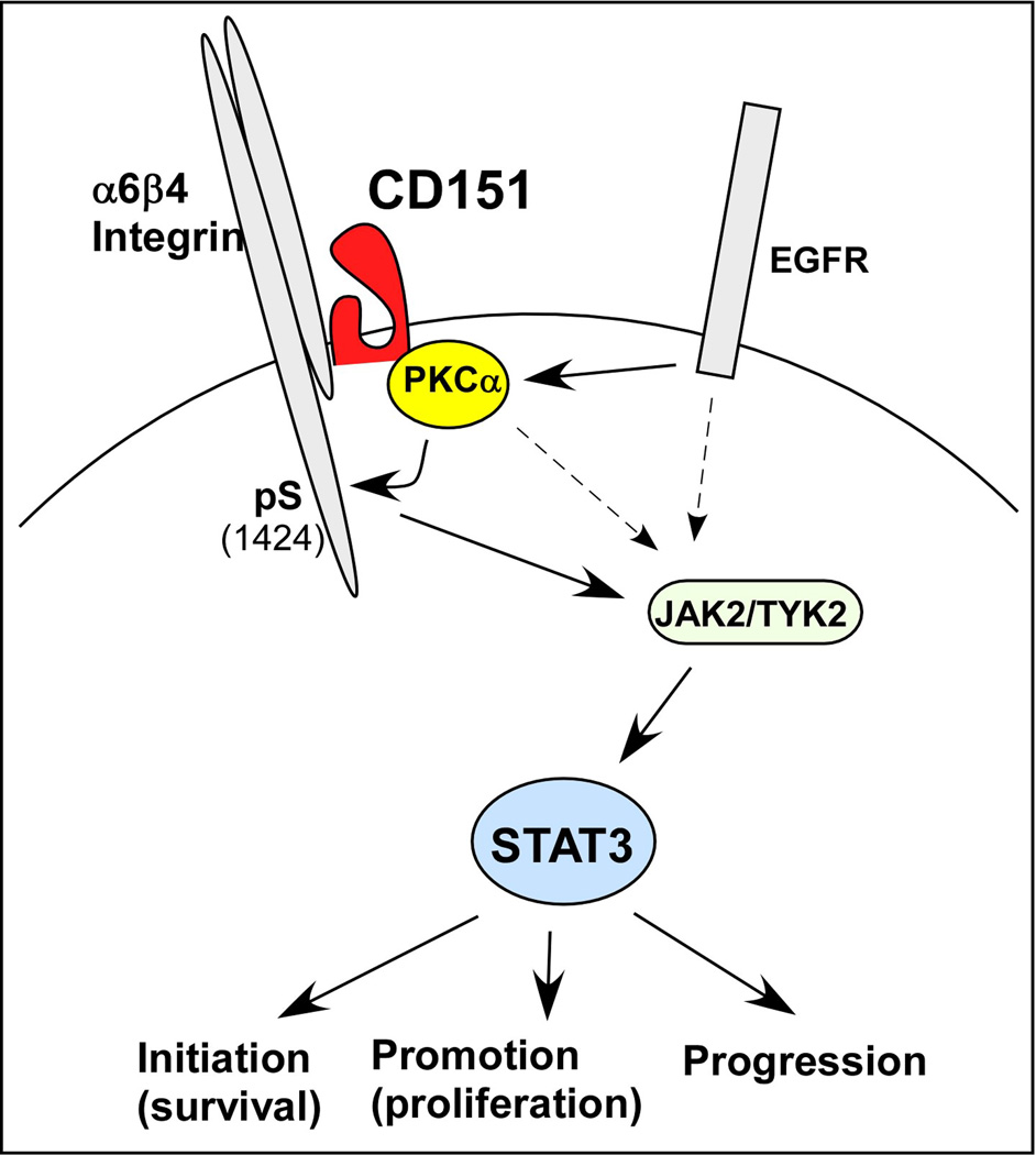
Here we provide the first evidence that tetraspanin CD151 can support de novo carcinogenesis. During two-stage mouse skin chemical carcinogenesis, CD151 reduces tumor lag time and increases incidence, multiplicity, size and progression to malignant squamous cell carcinoma (SCC), while supporting both cell survival during tumor initiation and cell proliferation during the promotion phase. In human skin SCC, CD151 expression is selectively elevated compared with other skin cancer types. CD151 support of keratinocyte survival and proliferation may depend on activation of transcription factor STAT3 (signal transducers and activators of transcription), a regulator of cell proliferation and apoptosis. CD151 also supports protein kinase C (PKC)α-α6β4 integrin association and PKC-dependent β4 S1424 phosphorylation, while regulating α6β4 distribution. CD151-PKCα effects on integrin β4 phosphorylation and subcellular localization are consistent with epithelial disruption to a less polarized, more invasive state. CD151 ablation, while minimally affecting normal cell and normal mouse functions, markedly sensitized mouse skin and epidermoid cells to chemicals/drugs including 7,12-dimethylbenz[α]anthracene (mutagen) and camptothecin (topoisomerase inhibitor), as well as to agents targeting epidermal growth factor receptor, PKC, Jak2/Tyk2 and STAT3. Hence, CD151 'co-targeting' may be therapeutically beneficial. These findings not only support CD151 as a potential tumor target, but also should apply to other cancers utilizing CD151/laminin-binding integrin complexes.


Conflict of interest statement

Conflict of Interest

The authors declare no competing financial interests related to the work described.

Figures

Figure 1
Figure 1
CD151 is elevated in human skin tissues. A, Staining of human skin SCC samples (5 left rows) and normal skin (right row). B, Enlarged CD151 staining of a human SCC sample. Red arrows indicate representative intracellular and peripheral CD151 staining. C, Immunohistochemical images show CD151 in normal skin (left panel) and skin SCC tumors with representative scoring = 1–4 (see Methods). D, Quantitation of results from a panel of different human skin cancer samples, such as shown in parts A and B. The right panel shows mean scores for 51 skin SCC and 32 combined BCC, MM and DFSP samples ± 95% C.I. *, P < 0.0001; Mann Whitney U test.
Figure 2
Figure 2
CD151 contribution to mouse skin tumor formation. A, Skin tumors in CD151+/+, +/−, and −/− mice 20 weeks after DMBA initiation. B, Delayed tumor appearance in CD151−/− mice. *, P < 0.0001 (log rank test). C, Average tumor numbers per mouse are indicated (± 95% CI). For data obtained after 20 weeks: **, P < 0.0001; *, P = 0.002 (Mann-Whitney U test). D, For each mouse group, tumor sizes are separated into three categories. Statistical analyses are based on two categories (Large; Medium+Small). For WT vs Null, P = 0.001; For Het vs Null, P = 0.009 (Fisher exact test).
Figure 3
Figure 3
DMBA-induced apoptosis in CD151 deficient mice. A, Representative active caspase-3 staining of epidermis from (a) untreated +/+, (b) +/+ mice with DMBA, (c) untreated −/−, and (d) −/− mice with DMBA. Caspase-3 positive cells appear in both hair follicle bulge regions (arrows) and interfollicular epidermis (arrowhead); Bar = 50 um. B, Quantitation of caspase-3 positive cells per unit area from data as in panel A. Shown are means ± SEM, n = 5; *, P < 0.001. C, Quantitation of keratinocyte apoptosis, after 4 hr of chemical treatment, based on accumulation of cytoplasmic histone-associated DNA fragments (see "Materials and methods"). Shown are means ± SEM, n = 5; *, P < 0.005; **, P < 0.006 (students t test)
Figure 4
Figure 4
TPA-induced epidermal proliferation. A, Mice (n = 5 per group) were treated with TPA and sacrificed 24 hrs after the last treatment. BrdU was injected 2 hr prior to sacrifice. H&E staining of epidermis from (a) untreated +/+, (b) +/+ treated with TPA, (c) untreated −/−, and (d) −/− mice treated with TPA. Bar = 50 um. B, Quantitation of epidermal thickness from wild type (black bar) and CD151 null mice (white bar). n = 5; *, P < 0.02. C, Immunohistochemical analysis of proliferation marker BrdU in dorsal skin after TPA treatment. D, Quantitation of BrdU positive cells in the epidermis of wild type (black bar) and CD151 null mice (white bar) after treatment with TPA. n = 5; *, P < 0.001
Figure 5
Figure 5
CD151 influences STAT3 activation. A, Mouse derived skin cell lines were lysed in 1% Triton X-100, and blotted for pSTAT3 (Tyr705, mAb from Cell Signaling Co.), total STAT3 (rabbit polyclonal Ab, Santa Cruz Co.), and β actin. B, CD151 was stably ablated in human A431 cells. Knockdown and control cells were treated with TPA (50 ng/ml) for indicated times, lysed, and then blotted as in part A. CD151 was detected using mAb 1A5. CD151 knockdown was >90% (bottom panel). Bar graphs, based on densitometry scans, show relative STAT3 activation C, Epidermis from +/+ and −/− mice was isolated 24 hr following DMBA treatment, and lysate proteins were blotted for activated STAT3, total STAT3 and β actin. Bar graphs show relative STAT3 activation. N = 5; *, P < 0.01. D, Epidermis from +/+ and −/− mice was isolated 24 hr following the last TPA treatment, and lysate proteins were blotted for activated STAT3, total STAT3 and β actin. Bar graphs show relative STAT3 activation. N = 5; *, P<0.02
Figure 6
Figure 6
CD151 and STAT3 inhibition effects. A, Equal numbers of freshly isolated primary mouse keratinocytes were cultured ± 5 µM nifuroxazide. After 2 days, cell proliferation was assessed using the MTT assay, which measures metabolic energy (left panel) and after 3 days total cell numbers were counted (right panel). Bars represent mean +/− SD for 3 independent experiments. *, P<0.05; **, P<0.001. B, STAT3 activation (pSTAT3-Y705) is shown for primary mouse keratinocytes treated for 2 days with 5 µM nifuroxazide. C, CD151 knockdown and control A431 cells were treated with nifuroxazide (2 µM or 5 µM) for indicated times, lysed, and then pSTAT3 (Y705) and total STAT3 were blotted, and ratios were determined from densitometric quantitation of blot intensities. See blots in supplemental Fig. S2A. D, A431 cells ± CD151 were treated with lapatinib, and then blotted for pSTAT3, total STAT3, pEGFR, and CD151 as indicated. Densitometric quantitation showed CD151 ablation causing pSTAT3/STAT3 ratios to decrease by 0%, 64%, and 84% (with 0, 1, 5 µM lapatinib, respectively). E, A341 cells ± CD151 were treated with STAT3 inhibitor ST3-01 (36), and then STAT3 activity was determined as indicated in supplemental Fig. S2B.
Figure 7
Figure 7
CD151 affects β4 distribution and phosphorylation. A, Mouse tumor-derived cell lines were permeabilized and stained for integrin β4. B, Keratinocytes were isolated from wild type and null mice and then cultured for a few weeks before integrin β4 subunit staining. Bar = 5 µm. (A, B) Linear staining intensity, across the diameter of representative cells, was measured using the Image J program (from NIH). C, Human A431 cells, stably ablated for CD151, were treated with TPA (50ng/ml) or EGF (100ng/ml) for 5 min and 1 hr, and then blotted for the indicated proteins, using antibodies for β4-S1424, β4-S1356, and total β4.
Figure 8
Figure 8
CD151 affects cPKC localization and function. A, A431 cells were stimulated with TPA (50ng/ml) or EGF (5 min and 1 hr), and then lysed in 1% Brij 56 detergent. PKCα and integrin β4 were immunoprecipitated (IP), followed by immunoblotting (IB) with antibodies to β4 and PKCα as indicated. WCL = whole cell lysates. *Numbers below the right panel represent mean density values for PKCα (associated with β4) from three independent experiments, including the one shown in the top right panel. B, A431 cells were stimulated with cPKC inhibitor Go 6976 for 2 hr, and then cells were lysed and blotted for pSTAT3, total STAT3, total PKC and CD151.
Figure 9
Figure 9
Scheme for CD151 contributions to chemical carcinogenesis in skin. Stimulation of epidermoid cells by EGF and TPA, through EGFR and PKCα, are enhanced when CD151 is present. Signaling is weakened when CD151 is absent, as evidenced by increased potency of agents inhibiting EGFR, PKCα, JAK2/TYK2, and STAT3. CD151 recruits PKC into proximity with β4 integrin. The β4 cytoplasmic tail has previously been linked to JAK2-STAT3 activation (24). It remains to be determined the extent to which CD151- and PKCα-dependent β4 S1424 phosphorylation directly contributes to STAT3 activation and skin oncogenesis. Results obtained here support previously demonstrated links of STAT3 to skin tumor initiation, promotion, and progression (10; 11).

Comment in

References

    1. Abel EL, Angel JM, Kiguchi K, DiGiovanni J. Nat Protoc. 2009;4:1350–1362. - PMC - PubMed
    1. Alam M, Ratner D. N Engl J Med. 2001;344:975–983. - PubMed
    1. Ang J, Lijovic M, Ashman LK, Kan K, Frauman AG. Cancer Epidemiol Biomarkers Prev. 2004;13:1717–1721. - PubMed
    1. Bachelder RE, Ribick MJ, Marchetti A, Falcioni R, Soddu S, Davis KR, Mercurio AM. J Cell Biol. 1999;147:1063–1072. - PMC - PubMed
    1. Baldwin G, Novitskaya V, Sadej R, Pochec E, Litynska A, Hartmann C, Williams J, Ashman L, Eble JA, Berditchevski F. J Biol Chem. 2008;283:35445–35454. - PubMed

Publication types

MeSH terms