. 2012 Jun 10;4(8):615-20.
doi: 10.1038/nchem.1375.

Crystal structure of Δ-[Ru(bpy)₂dppz]²⁺ bound to mismatched DNA reveals side-by-side metalloinsertion and intercalation

Affiliations

Crystal structure of Δ-[Ru(bpy)₂dppz]²⁺ bound to mismatched DNA reveals side-by-side metalloinsertion and intercalation

Hang Song et al. Nat Chem. .

Abstract

DNA mismatches represent a novel target in the development of diagnostics and therapeutics for cancer, because deficiencies in DNA mismatch repair are implicated in cancers, and cells that are repair-deficient show a high frequency of mismatches. Metal complexes with bulky intercalating ligands serve as probes for DNA mismatches. Here, we report the high-resolution (0.92 Å) crystal structure of the ruthenium 'light switch' complex Δ-[Ru(bpy)(2)dppz](2+) (bpy = 2,2'-bipyridine and dppz = dipyridophenazine), which is known to show luminescence on binding to duplex DNA, bound to both mismatched and well-matched sites in the oligonucleotide 5'-(dCGGAAATTACCG)(2)-3' (underline denotes AA mismatches). Two crystallographically independent views reveal that the complex binds mismatches through metalloinsertion, ejecting both mispaired adenosines. Additional ruthenium complexes are intercalated at well-matched sites, creating an array of complexes in the minor groove stabilized by stacking interactions between bpy ligands and extruded adenosines. This structure attests to the generality of metalloinsertion and metallointercalation as DNA binding modes.


Figures
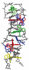
Figure 1
Figure 1
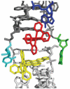
Structure of Δ-Ru(bpy)2dppz2+ (1) bound to the oligonucleotide 5′-C1G2G3A4A5A6T7T8A9C10C11G12-3′ shown in a front view (a) and rotated 90 degrees around the helix axis. Three DNA-binding modes are observed: (i) metalloinsertion, whereby the ruthenium complex (red) inserts the dppz ligand into the DNA duplex (gray) at the mismatched sites through the minor groove, extruding the mispaired adenosines (blue), (ii) metallointercalation, whereby the complex (green) binds between two well matched base pairs, and (iii) end-capping, whereby the complex (yellow) stacks with the terminal Watson-Crick pair of the duplex.
Figure 1
Figure 1
Structure of Δ-Ru(bpy)2dppz2+ (1) bound to the oligonucleotide 5′-C1G2G3A4A5A6T7T8A9C10C11G12-3′ shown in a front view (a) and rotated 90 degrees around the helix axis. Three DNA-binding modes are observed: (i) metalloinsertion, whereby the ruthenium complex (red) inserts the dppz ligand into the DNA duplex (gray) at the mismatched sites through the minor groove, extruding the mispaired adenosines (blue), (ii) metallointercalation, whereby the complex (green) binds between two well matched base pairs, and (iii) end-capping, whereby the complex (yellow) stacks with the terminal Watson-Crick pair of the duplex.
Figure 2
Figure 2
Two independent views of metalloinsertion at the mismatched sites. a, Superposition of the two independent views of metalloinsertion by the ruthenium complex at the mismatched sites, as viewed from the minor groove (A4-A9 site: ruthenium complex in red and DNA in gray, A9-A4 site: ruthenium complex in green and DNA in blue). The ruthenium complex inserts the dppz ligand from the minor groove and extrudes the mismatched adenosines, which are folded back into the minor groove. The two binding sites were superimposed using only the DNA backbone atoms (rmsd of 42 atoms = 0.607 Å). b, Superimposed metalloinsertion sites as viewed down the helical axis.
Figure 2
Figure 2
Two independent views of metalloinsertion at the mismatched sites. a, Superposition of the two independent views of metalloinsertion by the ruthenium complex at the mismatched sites, as viewed from the minor groove (A4-A9 site: ruthenium complex in red and DNA in gray, A9-A4 site: ruthenium complex in green and DNA in blue). The ruthenium complex inserts the dppz ligand from the minor groove and extrudes the mismatched adenosines, which are folded back into the minor groove. The two binding sites were superimposed using only the DNA backbone atoms (rmsd of 42 atoms = 0.607 Å). b, Superimposed metalloinsertion sites as viewed down the helical axis.
Figure 3
Figure 3
Two independent views of metallointercalation at well-matched sites. a, The ruthenium complex intercalates at the 5′-C1G2-3′ step through the dppz ligand (Ru in orange, DNA and bpy from a neighboring ruthenium in blue). b, Metallointercalation at the 5′-A6T7 -3′ step (Ru in magenta, DNA in gray).
Figure 3
Figure 3
Two independent views of metallointercalation at well-matched sites. a, The ruthenium complex intercalates at the 5′-C1G2-3′ step through the dppz ligand (Ru in orange, DNA and bpy from a neighboring ruthenium in blue). b, Metallointercalation at the 5′-A6T7 -3′ step (Ru in magenta, DNA in gray).
Figure 4
Figure 4
The end-capping complex. The duplex (dark gray) is end-capped by the ruthenium complex (red), which stacks between an extruded adenosine (blue) and the first complex (yellow) in a crystallographically related duplex (light gray). The last GC base pair (cytidine in cyan and guanosine in green) forms a frayed end.
Figure 5
Figure 5
Solution luminescence. Plot of integrated emission intensity (λex = 440 nm) of 1 μM Δ-Ru(bpy)2dppz2+ (1) with increasing concentration of Cu(phen)22+ in the presence of 12-mer mismatched (AA) and well-matched DNA (TA, 1 μM). Error bars indicate standard deviations in the measurements.

Comment in

Similar articles

Cited by

References

    1. Loeb LA. A mutator phenotype in cancer. Cancer Res. 2001;61:3230–3239. - PubMed
    1. Strauss BS. Frameshift mutation, microsatellites and mismatch repair. Mutat. Res. 1999;437:195–203. - PubMed
    1. Papadopoulos N, Lindblom A. Molecular basis of HNPCC: mutations of MMR genes. Hum. Mutat. 1997;10:89–99. - PubMed
    1. Peltomaki P. Deficient DNA mismatch repair: a common etiologic factor for colon cancer. Hum. Mol. Genet. 2001;10:735–740. - PubMed
    1. Lawes DA, SenGupta S, Boulos PB. The clinical importance and prognostic implications of microsatellite instability in sporadic cancer. Eur. J. Surg. Oncol. 2003;29:201–212. - PubMed

Publication types