. 2012 Sep-Oct;3(5):404-10.
doi: 10.4161/nucl.21222. Epub 2012 Jul 31.

Chromosomes without a 30-nm chromatin fiber

Affiliations

Chromosomes without a 30-nm chromatin fiber

Yasumasa Joti et al. Nucleus. 2012 Sep-Oct.

Abstract

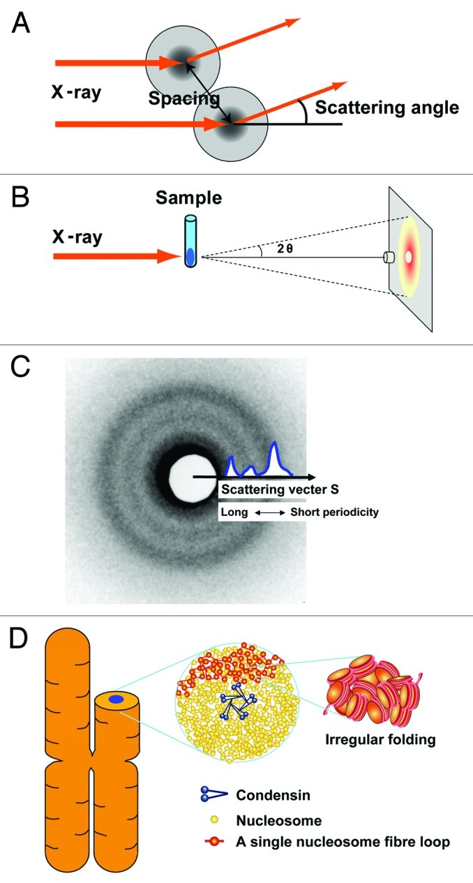
How is a long strand of genomic DNA packaged into a mitotic chromosome or nucleus? The nucleosome fiber (beads-on-a-string), in which DNA is wrapped around core histones, has long been assumed to be folded into a 30-nm chromatin fiber, and a further helically folded larger fiber. However, when frozen hydrated human mitotic cells were observed using cryoelectron microscopy, no higher-order structures that included 30-nm chromatin fibers were found. To investigate the bulk structure of mitotic chromosomes further, we performed small-angle X-ray scattering (SAXS), which can detect periodic structures in noncrystalline materials in solution. The results were striking: no structural feature larger than 11 nm was detected, even at a chromosome-diameter scale (~1 μm). We also found a similar scattering pattern in interphase nuclei of HeLa cells in the range up to ~275 nm. Our findings suggest a common structural feature in interphase and mitotic chromatins: compact and irregular folding of nucleosome fibers occurs without a 30-nm chromatin structure.


Figures

None
Figure 1. In the textbook model, a long DNA molecule is wrapped around a basic core histone octamer that consists of H2A, H2B, H3 and H4 histone proteins, and forms a nucleosome with a diameter of 11 nm. The nucleosome has long been assumed to be folded into 30-nm chromatin fibers before the higher-order organization of mitotic chromosomes or interphase nuclei occurs. We show a typical one-start helix model between two well-known structural models for 30-nm chromatin fibers: one-start helix (solenoid) and two-start helix (zigzag). The images are reproduced with minor modifications fromreference 60 with permission from Elsevier.
None
Figure 2. (A) When non-crystal materials are irradiated with X-rays, small-angle scattering generally reflects the size and spacing of internal structures. (B) Experimental setting: a chromosome pellet in a quartz capillary tube was exposed to a synchrotron X-ray beam and the scattering patterns were recorded with a CCD camera or imaging plate. The details of the setting are described in Nishino et al. (C) A typical scattering pattern is composed of concentric rings. The signals at smaller scattering angles [smaller size of the scattering vector (S) closer to the center] reflect larger periodic structures and vice versa. In Figure 3, the singles on the concentric rings are averaged and shown in a one-dimensional plot. The size of the scattering vector is defined by S = 2sin(θ)/λ, where λ is the wavelength and 2θ is the scattering angle. A periodic length is given by the inverse of S. Thus, "30-nm peak" refers to a scattering peak at S = 0.033 nm–1. (D) Chromosomes consist of irregularly folded nucleosome (beads on a string) fibers. Condensins (blue) hold the nucleosome fibers (red) globally around the chromosome center. Locally, the nucleosome fiber is folded in an irregular or disordered manner, forming loop structures that are collapsed toward the chromosome center (blue). The collapsed fiber (red) forms a domain that could be compatible with the large module observed by the Belmont group.
None
Figure 3. (A) SAXS profile of HeLa interphase nuclei, which were isolated using the Langmore and Paulson procedure., Three peaks at ~6, ~11 (faint) and ~30 nm were detected (arrows). In the plot of log(I × S2) vs. S, I is the measured average intensity and S is the size of the scattering vector, the inverse of the structure size or spacing (for details, see ref. 15). The 6- and 11-nm peaks are thought to come from edge-to-edge and face-to-face positioning of the nucleosomes, respectively. The 30-nm peak was assumed to represent the side-by-side packing of 30-nm chromatin fibers. (B) The presence and removal of ribosomes in the HeLa interphase nuclei were verified by western blotting. Nuclear lysates of ~2 × 10⁴, 1 × 10⁴, and 5 × 10³ cells were loaded into lanes 1–3, respectively. Proteins were separated by SDS-PAGE and then transferred to membranes. Ribosomal P-protein and histone H2B (control) and histone H3 (control) were detected using specific antibodies (H2B, Upstate 07–371; H3 Abcom ab1791). P-protein was detected in the original nuclei, but much less in the nuclei after washing, whereas histones H2B and H3 were similarly observed in both nuclei. (C) Only the 30-nm peak disappeared after removal of ribosome aggregates, whereas the other peaks remained.
None
Figure 4. (A) By ultrasmall-angle X-ray scattering (USAXS), no notable structures ~100–150 or 200–250 nm were detected in HeLa interphase nuclei. Beyond the ~275 nm range (red arrow), the slope in interphase chromatin decreased in magnitude. (B) For comparison, a USAXS profile of mitotic chromosomes is reproduced from Nishino et al. (C) The scattering intensity obeys the power law with respect to structure size or spacing. A plot of log (I) vs. log (S) on a straight line (red line) covers a wide range, extending over nearly three orders of magnitude. Least-squares fitting shows that I is proportional to S to the power of –3.36 (R = 0.999), suggesting that chromosomes do not possess notable regular structures over a very wide scale and exhibit a fractal nature of genome organization (see also ref. 15). (D) In the interphase nucleus, there are numerous compact chromatin domains like “chromatin liquid drops” (yellow balls). Red, transcribed nucleosome; Green, RNA polymerase and RNA. Formation of a 30-nm chromatin fiber might occur when nucleosome fibers are looped out from the chromatin domain or chromosome territory for transcription (top, see text). In our opinion, the transcriptional silencing can be established through dynamic capturing of transcriptional regions inside the compact chromatin domains. These domains can be considered as drops of viscous liquid, which could be formed by nucleosome-nucleosome interaction and a macromolecular crowding effect. Notably, this view is in line with predictions of the chromosome territory-interchromatin compartment (CT-IC) model, and previous evidence for an interchromatin compartment as well as the perichromatin region (see ref. 35).

Comment on

  • Nishino Y, Eltsov M, Joti Y, Ito K, Takata H, Takahashi Y, et al. Human mitotic chromosomes consist predominantly of irregularly folded nucleosome fibres without a 30-nm chromatin structure. EMBO J. 2012;31:1644–53. doi: 10.1038/emboj.2012.35.

References

    1. Alberts B, Johnson A, Lewis J, Raff M, Roberts K, Walter P. Molecular Biology of the Cell, Fifth Edition 2007.
    1. Kornberg RD, Lorch Y. Twenty-five years of the nucleosome, fundamental particle of the eukaryote chromosome. Cell. 1999;98:285–94. doi: 10.1016/S0092-8674(00)81958-3. - DOI - PubMed
    1. Olins DE, Olins AL. Chromatin history: our view from the bridge. Nat Rev Mol Cell Biol. 2003;4:809–14. - PubMed
    1. Robinson PJ, Rhodes D. Structure of the ‘30 nm’ chromatin fibre: a key role for the linker histone. Curr Opin Struct Biol. 2006;16:336–43. doi: 10.1016/j.sbi.2006.05.007. - DOI - PubMed
    1. Tremethick DJ. Higher-order structures of chromatin: the elusive 30 nm fiber. Cell. 2007;128:651–4. doi: 10.1016/j.cell.2007.02.008. - DOI - PubMed

Publication types

LinkOut - more resources