Review
. 2012 Aug;245(8):465-76.
doi: 10.1007/s00232-012-9464-0. Epub 2012 Jul 24.

Degradation of endocytosed gap junctions by autophagosomal and endo-/lysosomal pathways: a perspective

Affiliations
Review

Degradation of endocytosed gap junctions by autophagosomal and endo-/lysosomal pathways: a perspective

Matthias M Falk et al. J Membr Biol. 2012 Aug.

Abstract

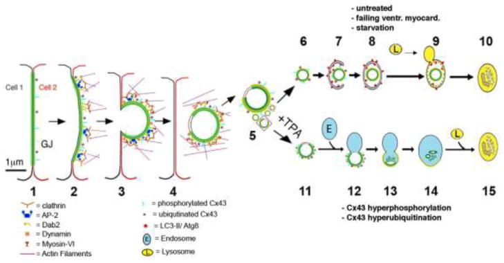
Gap junctions (GJs) are composed of tens to many thousands of double-membrane spanning GJ channels that cluster together to form densely packed channel arrays (termed GJ plaques) in apposing plasma membranes of neighboring cells. In addition to providing direct intercellular communication (GJIC, their hallmark function), GJs, based on their characteristic double-membrane-spanning configuration, likely also significantly contribute to physical cell-to-cell adhesion. Clearly, modulation (up-/down-regulation) of GJIC and of physical cell-to-cell adhesion is as vitally important as the basic ability of GJ formation itself. Others and we have previously described that GJs can be removed from the plasma membrane via the internalization of entire GJ plaques (or portions thereof) in a cellular process that resembles clathrin-mediated endocytosis. GJ endocytosis results in the formation of double-membrane vesicles [termed annular gap junctions (AGJs) or connexosomes] in the cytoplasm of one of the coupled cells. Four recent independent studies, consistent with earlier ultrastructural analyses, demonstrate the degradation of endocytosed AGJ vesicles via autophagy. However, in TPA-treated cells others report degradation of AGJs via the endo-/lysosomal degradation pathway. Here we summarize evidence that supports the concept that autophagy serves as the cellular default pathway for the degradation of internalized GJs. Furthermore, we highlight and discuss structural criteria that seem required for an alternate degradation via the endo-/lysosomal pathway.


Figures

Figure 1
Figure 1
Schematic representation of proposed steps that lead to GJ internalization (steps 1–3), cytoplasmic AGJ vesicle formation and fragmentation (steps 4, 5), and AGJ vesicle degradation by phago-/lysosomal (steps 6–10) and endo-/lysosomal pathways (steps 11–15) based on the previous work by others and us (see text for details). Note the proposed non-junctional membrane domains missing the green GJ label (shown in steps 4, 5, 11, 12), and the increased phosphorylation and ubiquitination on AGJ vesicles that fuse with endosomes (steps 11, 12 versus 6, 7). (Adapted from Falk et al. 2009 and Fong et al. 2012.)
Figure 2
Figure 2
Evidence for autophagic AGJ vesicle degradation. (A) HeLa cells were cotransfected with Cx43-mApple and the mammalian autophagy marker protein GFP-LC3 (left panel), or the activation-deficient LC3-mutant GFP-LC3(G120A) (right panel). In cells, a fraction of cytoplasmic LC3 (LC3-I) is covalently conjugated to phagophore-membranes (LC3-II) that localizes to autophagosomes; LC3(G120A) cannot be conjugated and remains cytoplasmic. Representative merged fluorescence images acquired 24 hours post transfections are shown. Individual and merged fluorescence signals of the boxed areas are shown below at higher magnification. Robust colocalization of cytoplasmic AGJ vesicles present in Cx43-mApple expressing cells (red puncta) with GFP-LC3-II (green puncta) was observed in GFP-LC3 expressing cells, but not in GFP-LC3(G120A) expressing cells. Representative colocalizing AGJ vesicles are marked with arrows; GJs are marked with arrowheads. Scale bars = 10 m. (B) Western blot analyses of total Cx43-GFP, or Cx43-YFP protein in transiently and stably expressing HeLa cells 72 hours after Beclin-1, a crucial autophagy-protein was depleted by RNAi-oligonucleotide transfection. SI-control cells were transfected with a scrambled non-targeting RISC-activating control oligonucleotide. Cx43-GFP/YFP was detected by probing with polyclonal anti-Cx43 antibodies. Normalized quantitative analyses revealed a two to three-fold accumulation of Cx43-GFP/YFP protein in Beclin-1-KD over RNAi-control cells. (C) Quantitative AGJ vesicle analyses performed 72 hours post RNA oligonucleotide-transfection indicated a significant cytoplasmic AGJ accumulation in Beclin-1/(Atg6), LAMP-2, and p62/SQSTM1 (all autophagy-related proteins) depleted cells (≥ 50%, marked with asterisks) compared to RNAi-control cells (panel 1, left). Less pronounced AGJ vesicle-accumulation was observed in LC3-depleted cells (32%), and this was attributed to sufficient inactive LC3-I that may have remained in the LC3-KD cells and may have been converted into active LC3-II. In addition, quantitative analyses of AGJ vesicles (panel 2, center left), LC3-positive autophagosomes (panel 3, center right), and colocalizing AGJ/autophagosomes (panel 4, right) in Beclin-1 and p62/SQSTM1 knockdown cells revealed a significant increase of AGJs, a significant decrease of autophagosomes, and significantly reduced AGJ/autophagosome colocalization in three independent experiments (** = p<0.01; *** = p<0.001). (D) Multiple stages characteristic of progressive autophagosome formation and maturation that formed around AGJ vesicles revealed by ultrastructural analyses of Cx43-GFP expressing HeLa cell preparations. Double-membrane cisternae (presumably isolation membranes, marked with arrows) progressively encircled AGJ vesicles (panels a–c), coalesced into phagophores (panels c–e) and fused with lysosomes (L, panels d, e), resulting in AGJ degradation inside the phagosome (panel f). (Figure portions A to D are reproduced with permission from Fong et al. 2012.) (E–H) Autophagosomes presumably degrading endogenous AGJs in vivo, identified in the equine stratum spinosum (E, reproduced with permission from Leach and Oliphant 1984); rat liver (F, reproduced with permission from Pfeifer 1980); mouse embryonic fibroblasts (G, reproduced with permission from Lichtenstein et al. 2011); and mammalian cardiomyocytes (H, reproduced with permission from Severs et al. 1989). Scale bars = 100 nm
Figure 3
Figure 3
Fine structure and composition of AGJ vesicles. (A) Double-membrane AGJ vesicles can contain a patch of non-junctional membrane where the two membrane layers are separated (enlarged in inserts). These non-junctional membrane patches appear to be derived from plasma membrane that was located immediately adjacent to the GJ plaque and was concomitantly endocytosed. (B) These non-junctional AGJ membrane domains label with extracellularly applied, fluorescence-labeled wheat germ agglutinin (WGA) (red puncta on green AGJ vesicles marked with arrows) in Cx43-YFP expressing cells. Stable Cx43-YFP expressing HeLa cells were incubated for 2–4 h with Alexa594-labelled WGA and examined by fluorescence microscopy. Low magnification survey image (panel 1, left) and high-resolution images of internalized AGJ vesicles (panels 2–5, right) are shown (scale bars = nm in [A], and m in [B]). (Figure 3A is adapted from (Piehl et al. 2007).)

References

    1. Baker SM, Kim N, Gumpert AM, Segretain D, Falk MM. Acute internalization of gap junctions in vascular endothelial cells in response to inflammatory mediator-induced G-protein coupled receptor activation. FEBS Lett. 2008;582:4039–4046. - PMC - PubMed
    1. Baldwin AL, Thurston G. Mechanics of endothelial cell architecture and vascular permeability. Crit Rev Biomed Eng. 2001;29:247–278. - PubMed
    1. Barriere H, Nemes C, Lechardeur D, Khan-Mohammad M, Fruh K, Lukacs GL. Molecular basis of oligoubiquitin-dependent internalization of membrane proteins in Mammalian cells. Traffic. 2006;7:282–297. - PubMed
    1. Beardslee MA, Laing JG, Beyer EC, Saffitz JE. Rapid turnover of connexin43 in the adult rat heart. Circ Res. 1998;83:629–635. - PubMed
    1. Bejarano E, Girao H, Yuste A, Patel B, Marques C, Spray DC, Pereira P, Cuervo AM. Autophagy modulates dynamics of connexins at the plasma membrane in a ubiquitin-dependent manner. Mol Biol Cell. 2012;23:2156–2169. - PMC - PubMed

Publication types

LinkOut - more resources