. 2012 Aug 31;287(36):30789-99.
doi: 10.1074/jbc.M112.390120. Epub 2012 Jul 23.

DNA damage-induced heterogeneous nuclear ribonucleoprotein K sumoylation regulates p53 transcriptional activation

Affiliations

DNA damage-induced heterogeneous nuclear ribonucleoprotein K sumoylation regulates p53 transcriptional activation

Federico Pelisch et al. J Biol Chem. .

Abstract

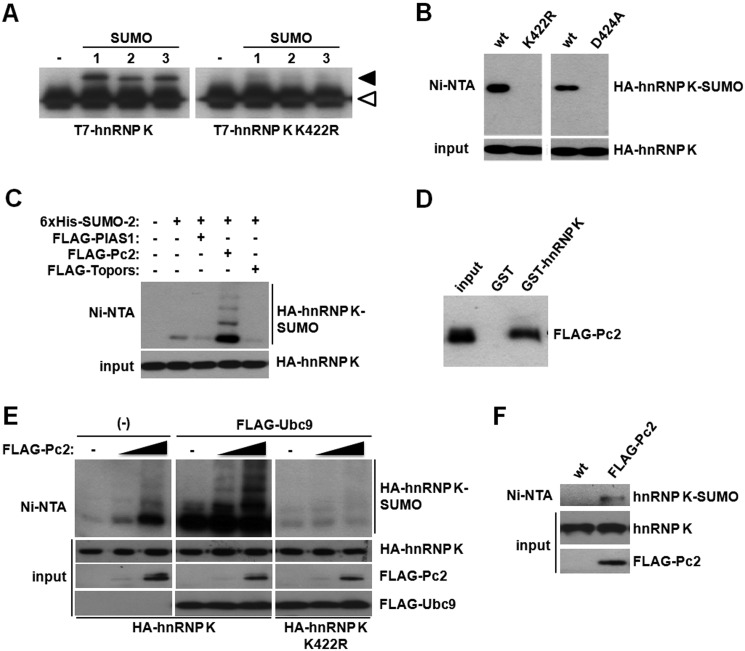
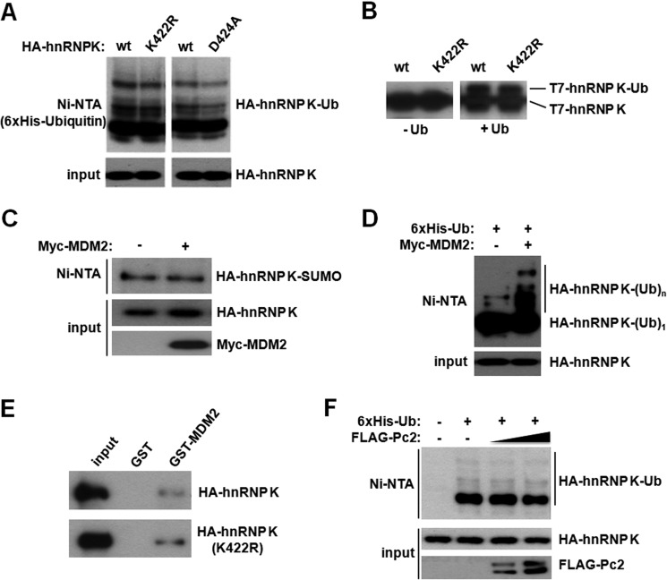
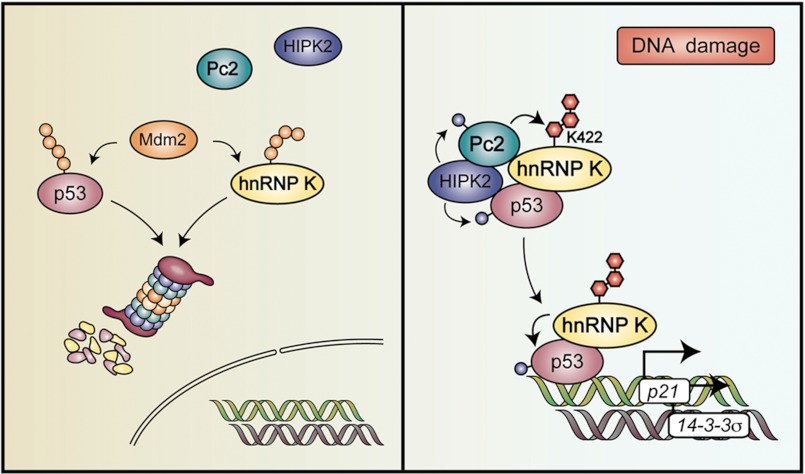
Heterogeneous nuclear ribonucleoprotein (hnRNP) K is a nucleocytoplasmic shuttling protein that is a key player in the p53-triggered DNA damage response, acting as a cofactor for p53 in response to DNA damage. hnRNP K is a substrate of the ubiquitin E3 ligase MDM2 and, upon DNA damage, is de-ubiquitylated. In sharp contrast with the role and consequences of the other post-translational modifications, nothing is known about the role of SUMO conjugation to hnRNP K in p53 transcriptional co-activation. In the present work, we show that hnRNP K is modified by SUMO in lysine 422 within its KH3 domain, and sumoylation is regulated by the E3 ligase Pc2/CBX4. Most interestingly, DNA damage stimulates hnRNP K sumoylation through Pc2 E3 activity, and this modification is required for p53 transcriptional activation. Abrogation of hnRNP K sumoylation leads to an aberrant regulation of the p53 target gene p21. Our findings link the DNA damage-induced Pc2 activation to the p53 transcriptional co-activation through hnRNP K sumoylation.


Figures

FIGURE 1.
FIGURE 1.
HnRNP K is a SUMO substrate in cells and in vitro. A, summary of the highest probability site according to both SUMOplot and SUMOsp, lysine 422 (top), and the schematic of hnRNP K highlighting its lysine 422 residing within the KH3 domain (bottom). B, HEK293T cells were transfected with an HA-tagged hnRNP K expression vector either with or without SUMO-2. An aliquot of cells was taken as input, and the remainder was subject to denaturing Ni2+ affinity chromatography. Both fractions were analyzed by Western blot with an anti-HA antibody. C, for the analysis of endogenous hnRNP K sumoylation, cell lysates were analyzed as in B, although an anti-hnRNP K antibody was used for the Western blot. D, pull-down assay was performed using HEK293T lysates expressing HA-hnRNP K. Samples were analyzed by Western blot with an anti-HA antibody. E, purified recombinant T7-hnRNP K was incubated with Aos1-Uba2, increasing amounts of Ubc9, and SUMO-2. Reactions were stopped by addition of 2× Laemmli sample buffer and analyzed by Western blot with an anti-T7 antibody. F, in vitro reactions as in E were performed in triplicate, and the signal was quantified with ImageJ after corroboration that all the bands in the developed films were in a linear range. Data are presented as mean ± S.E.
FIGURE 2.
FIGURE 2.
The polycomb protein Pc2 exerts E3 ligase activity toward hnRNP K. A, in vitro sumoylation reactions were performed using SUMO-1, -2, or -3 together with wild type or the K422R mutant T7-tagged hnRNP K. The white arrowhead denotes the migration of non-sumoylated T7-hnRNP K, while the black arrowhead indicates the migration of sumoylated T7-hnRNP K. B, HEK293T cells were transfected with wild type, K422R, or D424A mutant HA-tagged hnRNP K together with His6-tagged SUMO-2. An aliquot of cells was taken as input, and the remainder was subject to denaturing Ni2+ affinity chromatography. Both fractions were analyzed by Western blot with an anti-HA antibody. C, HEK293T cells were transfected with the indicated expression vectors and analyzed as above. D, pull-down assay was performed using HEK293T lysates expressing FLAG-Pc2. Samples were analyzed by Western blot with an anti-FLAG antibody. E, HEK293T cells were transfected with the indicated expression vectors and analyzed as above. F, HEK293T cells were transfected with His6-tagged SUMO-2 either with or without Pc2 and endogenous hnRNP K sumoylation was analyzed.
FIGURE 3.
FIGURE 3.
Lysine 422 is conjugated by SUMO and not ubiquitin. A, HEK293T cells were transfected either with wild type, K422R, or D424A HA-tagged hnRNP K expression vectors, together with His6-tagged ubiquitin, as indicated. Cells were subject to denaturing nickel affinity chromatography as above and analyzed by Western blot with an anti-HA antibody. B, recombinant T7-hnRNP K and T7-hnRNP K K422R were ubiquitylated in vitro in the presence of Uba1, UbcH5b, and MDM2. The left panel corresponds to reactions lacking ubiquitin. C, cells were transfected with the indicated plasmids and analyzed as in A. D, cells were transfected with HA-tagged hnRNP K together with His6-tagged ubiquitin, and 48 h later they were treated with 10 μm MG132 for 5 h before lysis and Ni2+ affinity chromatography as above. E, lysates expressing HA-hnRNP K or the K422R mutant were incubated either with GST or GST-MDM2, and the pull-down assay was performed as described under “Experimental Procedures.” Samples were analyzed by Western blot with an anti-HA antibody. F, cells were transfected with the indicated plasmids and analyzed by Ni2+ affinity chromatography as in D.
FIGURE 4.
FIGURE 4.
hnRNP K sumoylation is stimulated by DNA damage. A, HCT-116 cells were transfected with HA-tagged hnRNP K together with His6-tagged SUMO-2, and 48 h later they were left untreated or treated with 1 μm doxorubicin, 1 μm Etoposide, or UV light (40 J/m2) for 4 h. SUMO conjugation to hnRNP K was analyzed by Ni2+ affinity chromatography as above. B, doxorubicin-induced hnRNP K sumoylation depends on Lys-422. Cells were transfected and processed as above. C, endogenous hnRNP K sumoylation is induced by doxorubicin. D, cells were transfected with His6-tagged hnRNP K, and 48 h later they were treated with doxorubicin and hnRNP K was purified by Ni2+ affinity chromatography and endogenous SUMO2/3 conjugation was assessed by Western blot. E, cells were transfected with HA-tagged hnRNP K together with His6-tagged ubiquitin; 48 h later they were treated with 1 μm doxorubicin for 3 h in the presence of 10 μm MG132 and ubiquitin conjugation to hnRNP K was analyzed by Ni2+ affinity chromatography as above.
FIGURE 5.
FIGURE 5.
hnRNP K sumoylation is required for p53 transcriptional co-activation. A, HEK 293T cells were transfected with the indicated plasmids and then analyzed by Ni2+ affinity chromatography. B and C, Pc2 and HIPK2 regulate hnRNP sumoylation. HEK 293T cells were transfected with the indicated plasmids, and 48 h later they were treated with 1 μm doxorubicin (Dox) for 4 h. Then, HA-hnRNP K sumoylation was analyzed by Ni2+ affinity chromatography. D, cells were transfected with a plasmid carrying 16 tandem p53 binding sites upstream of the firefly luciferase coding sequence, together with HA-hnRNP K, Pc2, or HIPK2 and lysed 48 h later for luciferase activity measurement. E, cells were transfected with a plasmid carrying 16 tandem p53 binding sites upstream of the firefly luciferase coding sequence, together with HA-hnRNP K, Pc2 ΔSIM, or HIPK2 K221A and analyzed as in D. Data are represented as mean ± S.E., and p values of Student's t test are shown.
FIGURE 6.
FIGURE 6.
hnRNP K sumoylation is required for p53 transcriptional co-activation. Pc2-mediated sumoylation of hnRNP K at lysine 422 activates p53-dependent transcription. A, HCT-116 cells were transfected with a plasmid carrying 16 tandem p53 binding sites upstream of the firefly luciferase coding sequence, together with HA-hnRNP K, or HA-hnRNP K K422R and FLAG-Pc2. Data are represented as mean ± S.E. B, HCT-116 cells were transfected with a plasmid carrying 16 tandem p53 binding sites upstream of the firefly luciferase coding sequence, together with HA-hnRNP K, or HA-hnRNP K K422R and FLAG-HIPK2. C, hnRNP K is required for proper p21 expression upon DNA damage. HCT-116 cells were transfected either with a control (Ctl) or an hnRNP K siRNA and 48 hs later they were treated with 1 μm doxorubicin (Dox) for 24 h before RNA extraction and real-time PCR analysis. D, HCT-116 cells were transfected either with a control (Ctl.) or an hnRNP K siRNA, and 24 h later they were transfected with the indicated hnRNP K construct. The next day, cells were treated with 1 μm doxorubicin (Dox) for 24 h before RNA extraction and real-time PCR analysis as in C. Real-time PCR experiments were always performed in triplicate, and data are represented as mean ± S.E. In parallel, HA-hnRNP K and β-actin expression was monitored by Western blot. All HA (upper) and β-actin (lower) panels correspond to cropped images of the same blot. Luciferase reporter assays were performed in duplicate and real-time PCR experiments in triplicate. p values of the Student's t test are shown.
FIGURE 7.
FIGURE 7.
p53 is required for hnRNP K transcriptional co-activation. hnRNP K and Pc2 activate p53 target gene expression synergistically. Wild type (p53 +/+) and p53-null (p53 −/−) HCT-116 cells were transfected with the indicated plasmids, and RNA was extracted 48 h later. p21 (A and B) and 14-3-3σ (C and D) mRNA levels were quantified by real-time PCR. In all cases, HSPCB was used as a housekeeping gene. Real-time PCR experiments were performed in triplicates. E, HCT-116 p53 −/− cells were transfected with a p21 reporter plasmid together with hnRNP K or hnRNP K D424A, and a p53 expression vector. Data are represented as mean ± S.E. p values of the Student's t test are shown.
FIGURE 8.
FIGURE 8.
Model illustrating the role of hnRNP K sumoylation upon DNA damage. Under normal conditions, p53 and hnRNP K are ubiquitylated and degraded by the proteasome. Upon treatment with DNA-damaging agents, HIPK2 phosphorylates and activates Pc2, which in turn, leads to hnRNP K sumoylation. Sumoylated hnRNP K then serves as a cofactor for p53-mediated gene expression. The model incorporates the new data obtained in the present report with data from previous studies.

References

    1. Geiss-Friedlander R., Melchior F. (2007) Concepts in sumoylation: a decade on. Nat. Rev. Mol. Cell Biol. 8, 947–956 - PubMed
    1. Hay R. T. (2005) SUMO: a history of modification. Mol. Cell 18, 1–12 - PubMed
    1. Bernassola F., Karin M., Ciechanover A., Melino G. (2008) The HECT family of E3 ubiquitin ligases: multiple players in cancer development. Cancer Cell 14, 10–21 - PubMed
    1. Deshaies R. J., Joazeiro C. A. (2009) RING domain E3 ubiquitin ligases. Annu. Rev. Biochem. 78, 399–434 - PubMed
    1. Kahyo T., Nishida T., Yasuda H. (2001) Involvement of PIAS1 in the sumoylation of tumor suppressor p53. Mol. Cell 8, 713–718 - PubMed

Publication types

MeSH terms