. 2012 Jul 18:3:209.
doi: 10.3389/fimmu.2012.00209. eCollection 2012.

Autoimmunity in wiskott-Aldrich syndrome: an unsolved enigma

Affiliations

Autoimmunity in wiskott-Aldrich syndrome: an unsolved enigma

Marco Catucci et al. Front Immunol. .

Abstract

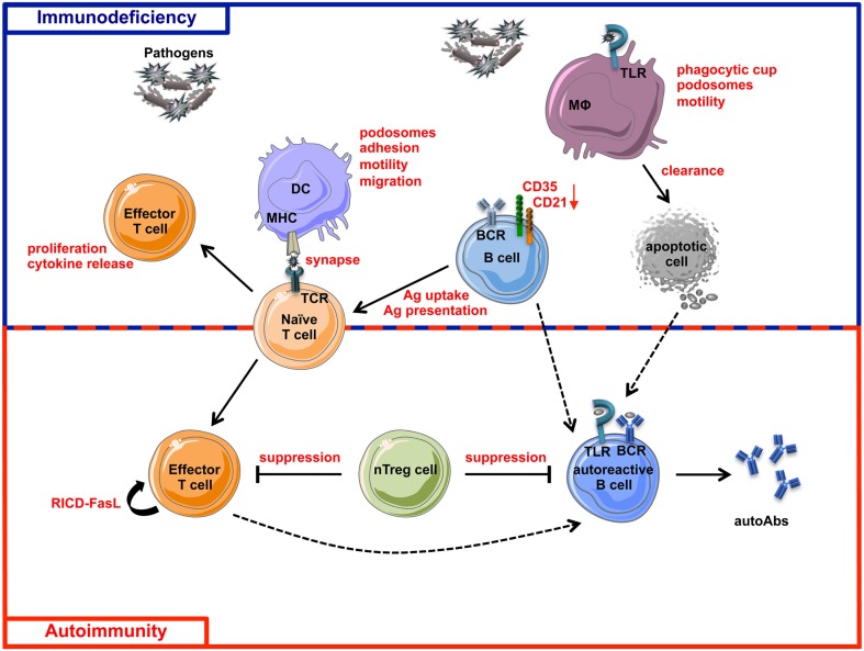
Wiskott-Aldrich Syndrome (WAS) is a severe X-linked Primary Immunodeficiency that affects 1-10 out of 1 million male individuals. WAS is caused by mutations in the WAS Protein (WASP) expressing gene that leads to the absent or reduced expression of the protein. WASP is a cytoplasmic protein that regulates the formation of actin filaments in hematopoietic cells. WASP deficiency causes many immune cell defects both in humans and in the WAS murine model, the Was(-/-) mouse. Both cellular and humoral immune defects in WAS patients contribute to the onset of severe clinical manifestations, in particular microthrombocytopenia, eczema, recurrent infections, and a high susceptibility to develop autoimmunity and malignancies. Autoimmune diseases affect from 22 to 72% of WAS patients and the most common manifestation is autoimmune hemolytic anemia, followed by vasculitis, arthritis, neutropenia, inflammatory bowel disease, and IgA nephropathy. Many groups have widely explored immune cell functionality in WAS partially explaining how cellular defects may lead to pathology. However, the mechanisms underlying the occurrence of autoimmune manifestations have not been clearly described yet. In the present review, we report the most recent progresses in the study of immune cell function in WAS that have started to unveil the mechanisms contributing to autoimmune complications in WAS patients.

Keywords: B lymphocytes; T lymphocytes; Wiskott–Aldrich syndrome; autoimmunity; primary immunodeficiency.


Figures

Figure 1
Figure 1
Wiskott–Aldrich syndrome structure and interacting proteins. TCR, BCR, chemokine receptors, TLRs, integrins, and the Fc receptor γ-chain can promote the release of GDP from Rho family GTPases, allowing GTP to bind. In immune cells, the major Rho GTPase is the Cell Division Cycle 42 (CDC42). The WASP-Homology 1 (WH1) domain mediates the binding to WASP-Interacting Protein (WIP; Ramesh et al., 1997). The Phosphatidylinositol-4,5-bisphosphate (PIP2) links to the Basic (B) domain and stabilizes WASP active form. The binding of the GTPase-Binding Domain (GBD) with CDC42 induces WASP activation (Kolluri et al., ; Symons et al., ; Miki et al., 1998). The proline-rich region (PPP) provides binding sites for the Vasodilator-Stimulated Phosphoprotein (VASP), and also for SRC family tyrosine kinases and SRC Homology 3 (SH3) domain-containing proteins such as the adaptor proteins GRB2, FYN, PI3K, and NCK. The Verprolin-homology (V) domain binds to actin monomers, and the Cofilin-homology (C) and Acidic (A) domains bind to the Actin-Related Protein (ARP)2/3 complex. The V/C/A region functions as the platform to initiate actin polymerization (Park et al., 2010).
Figure 2
Figure 2
Schematic view of cellular defects described in WASP-deficient cells. MΦ, Macrophage; Neut., Neutrophil.
Figure 3
Figure 3
Role of WASP in TCR and BCR signaling pathways. The main signaling molecules (blue and green ovals) acting downstream of TCR (A), BCR, and TLRs (B) are depicted. The red boxes indicate the main pathways induced downstream of these receptors. The role of WASP in these pathways is indicated by stars close to interacting molecules: red stars show the interactions that are demonstrated, whereas the blue stars with question marks show hypothetical involvement of WASP in TLR signaling pathways.
Figure 4
Figure 4
Schematic view of immunodeficiency and autoimmunity in WAS. The impairment of both innate and adaptive immune systems is responsible of immunodeficiency (blue box) and autoimmunity (red box) in WAS. Immune cell defects described in WASP-deficient cells are shown in red. The incomplete pathogen clearance is sustained by cytoskeleton and functional defects of macrophages, DCs, T cells, B cells, and their defective interactions. The reduced expression of CD21 and CD35, two complement receptors involved in antigen uptake and presentation and also in negative selection of self-reactive B cells, places B cells at the interface between immunodeficiency and autoimmunity. Defective suppression of WASP-deficient nTreg cells toward both T and B cells contributes to the tolerance breakdown in WAS. Defect in RICD process, resulting in defective effector T cell apoptotic death after TCR restimulation, concurs in the persistence of T cell response to pathogens or self-antigens. Additionally, intrinsic B cell defects contribute to autoimmunity in WAS, probably via a TLR-mediated mechanism. Dashed lines represent hypothetical mechanisms involved in WAS-related autoimmunity. MHC, Major Histocompatibility Complex; RICD, Restimulation-Induced Cell Death; autoAbs, autoantibodies; Ag, Antigen.

References

    1. Abdul-Manan N., Aghazadeh B., Liu G. A., Majumdar A., Ouerfelli O., Siminovitch K. A., Rosen M. K. (1999). Structure of Cdc42 in complex with the GTPase-binding domain of the ‘Wiskott-Aldrich syndrome’ protein. Nature 399, 379–38310.1038/20726 - DOI - PubMed
    1. Adriani M., Aoki J., Horai R., Thornton A. M., Konno A., Kirby M., Anderson S. M., Siegel R. M., Candotti F., Schwartzberg P. L. (2007). Impaired in vitro regulatory T cell function associated with Wiskott-Aldrich syndrome. Clin. Immunol. 124, 41–4810.1016/j.clim.2007.02.001 - DOI - PMC - PubMed
    1. Adriani M., Jones K. A., Uchiyama T., Kirby M. R., Silvin C., Anderson S. M., Candotti F. (2011). Defective inhibition of B-cell proliferation by Wiskott-Aldrich syndrome protein-deficient regulatory T cells. Blood 117, 6608–661110.1182/blood-2010-12-322834 - DOI - PMC - PubMed
    1. Albert M. H., Bittner T. C., Nonoyama S., Notarangelo L. D., Burns S., Imai K., Espanol T., Fasth A., Pellier I., Strauss G., Morio T., Gathmann B., Noordzij J. G., Fillat C., Hoenig M., Nathrath M., Meindl A., Pagel P., Wintergerst U., Fischer A., Thrasher A. J., Belohradsky B. H., Ochs H. D. (2010). X-linked thrombocytopenia (XLT) due to WAS mutations: clinical characteristics, long-term outcome, and treatment options. Blood 115, 3231–323810.1182/blood-2009-09-239087 - DOI - PubMed
    1. Antoine C., Muller S., Cant A., Cavazzana-Calvo M., Veys P., Vossen J., Fasth A., Heilmann C., Wulffraat N., Seger R., Blanche S., Friedrich W., Abinun M., Davies G., Bredius R., Schulz A., Landais P., Fischer A. (2003). Long-term survival and transplantation of haemopoietic stem cells for immunodeficiencies: report of the European experience 1968-99. Lancet 361, 553–56010.1016/S0140-6736(03)12513-5 - DOI - PubMed