. 2012 Jul 3;103(1):38-47.
doi: 10.1016/j.bpj.2012.05.039.

Dynamic force spectroscopy on supported lipid bilayers: effect of temperature and sample preparation

Affiliations

Dynamic force spectroscopy on supported lipid bilayers: effect of temperature and sample preparation

Andrea Alessandrini et al. Biophys J. .

Abstract

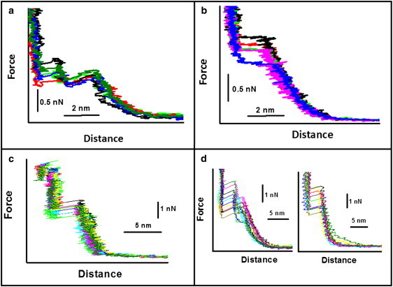
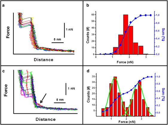
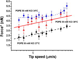
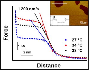
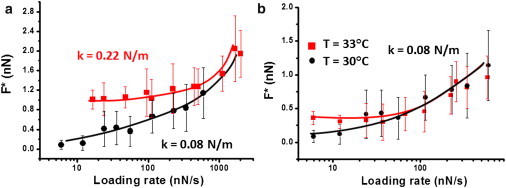
Biological membranes are constantly exposed to forces. The stress-strain relation in membranes determines the behavior of many integral membrane proteins or other membrane related-proteins that show a mechanosensitive behavior. Here, we studied by force spectroscopy the behavior of supported lipid bilayers (SLBs) subjected to forces perpendicular to their plane. We measured the lipid bilayer mechanical properties and the force required for the punch-through event characteristic of atomic force spectroscopy on SLBs as a function of the interleaflet coupling. We found that for an uncoupled bilayer, the overall tip penetration occurs sequentially through the two leaflets, giving rise to two penetration events. In the case of a bilayer with coupled leaflets, penetration of the atomic force microscope tip always occurred in a single step. Considering the dependence of the jump-through force value on the tip speed, we also studied the process in the context of dynamic force spectroscopy (DFS). We performed DFS experiments by changing the temperature and cantilever spring constant, and analyzed the results in the context of the developed theories for DFS. We found that experiments performed at different temperatures and with different cantilever spring constants enabled a more effective comparison of experimental data with theory in comparison with previously published data.


Figures

Figure 1
Figure 1
(a) Force curves obtained on an uncoupled supported POPE bilayer prepared at 15°C. (b) Force curves obtained on a coupled POPE supported bilayer prepared at 30°C. (c) Force curves obtained on an uncoupled POPG bilayer prepared at 15°C. (d) Force curves obtained on a POPG bilayer prepared at 20°C for tip speeds of 359 nm/s (left) and 930 nm/s (right). Cantilever spring constant: 0.08 ± 0.2 N/m.
Figure 2
Figure 2
(a) Force curves obtained on a POPG bilayer at 27°C and tip speed of 598 nm/s. (b) Jump-through force values distribution corresponding to the case in panel a. (c) Force curves obtained on the same POPG bilayer as in panel a, but at 24°C. The arrow points to the region of the force curves in which the electrostatic interaction is increased. (d) Jump-through force values distribution corresponding to the case in panel c.
Figure 3
Figure 3
Most probable jump-through force as a function of tip speed for a POPE bilayer with coupled leaflets. The three sequences of data were obtained at 27° (squares) (on liquid-disordered domains in the phase transition region), at 34°C (circles) and 38°C (triangles). The dotted line for the data at 34°C is a guide for the eye, whereas the continuous and dashed lines are linear fits to the data. The lines are guides to the eye. Spring constant of the cantilever: 0.07 ± 0.02 N/m.
Figure 4
Figure 4
Force curves obtained on a POPE supported bilayer as a function of temperature at a constant tip speed of 1200 nm/s. Each reported curve is an average of >200 force curves. The overlaid lines highlight the steepness of the contact region between the tip and the bilayer as an indication of the bilayer deformability. Inset: AFM image of a POPE supported bilayer with a defect that allows one to measure the bilayer height. The cross section shown in the lower part of the inset corresponds to the horizontal line shown in the image.
Figure 5
Figure 5
(a) Most probable jump-through force as a function of the loading rate on a POPE bilayer obtained with two cantilevers of different spring constants (squares: k = 0.22 N/m; circles: k = 0.08 N/m) (T = 30°C). (b) Most probable jump-through force as a function of the loading rate on the same POPE bilayer in panel a, obtained at two different temperatures (spring constant = 0.08 N/m). All of the continuous lines are guides to the eye.

References

    1. Sackmann E. Supported membranes: scientific and practical applications. Science. 1996;271:43–48. - PubMed
    1. Tamm L.K., McConnell H.M. Supported phospholipid bilayers. Biophys. J. 1985;47:105–113. - PMC - PubMed
    1. Alessandrini A., Facci P. Unraveling lipid/protein interaction in model lipid bilayers by atomic force microscopy. J. Mol. Recognit. 2011;24:387–396. - PubMed
    1. Alessandrini A., Seeger H.M., Facci P. What do we really measure in AFM punch-through experiments on supported lipid bilayers? Soft Matter. 2011;7:7054–7064.
    1. Alessandrini A., Facci P. AFM: a versatile tool in biophysics. Meas. Sci. Technol. 2005;16:R65–R92.

Publication types

MeSH terms

LinkOut - more resources