

NADH distribution in live progenitor stem cells by phasor-fluorescence lifetime image microscopy
- PMID: 22828352
- PMCID: PMC3388207
- DOI: 10.1016/j.bpj.2012.05.038
NADH distribution in live progenitor stem cells by phasor-fluorescence lifetime image microscopy
Abstract
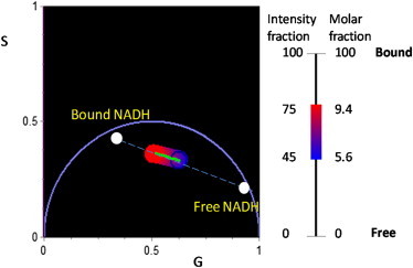
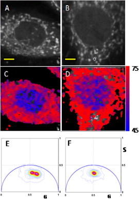
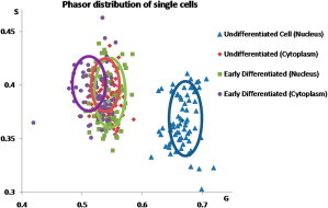
NADH is a naturally fluorescent metabolite associated with cellular respiration. Exploiting the different fluorescence lifetime of free and bound NADH has the potential to quantify the relative amount of bound and free NADH, enhancing understanding of cellular processes including apoptosis, cancer pathology, and enzyme kinetics. We use the phasor-fluorescence lifetime image microscopy approach to spatially map NADH in both the free and bound forms of live undifferentiated and differentiated myoblast cells. The phasor approach graphically depicts the change in lifetime at a pixel level without the requirement for fitting the decay. Comparison of the spatial distribution of NADH in the nucleus of cells induced to differentiate through serum starvation and undifferentiated cells show differing distributions of bound and free NADH. Undifferentiated cells displayed a short lifetime indicative of free NADH in the nucleus and a longer lifetime attributed to the presence of bound NADH outside of the nucleus. Differentiating cells displayed redistribution of free NADH with decreased relative concentration of free NADH within the nucleus whereas the majority of NADH was found in the cytoplasm.
Copyright © 2012 Biophysical Society. Published by Elsevier Inc. All rights reserved.
Figures
References
-
- White, B. P., F. J. McDonald, and L. Willmott. 2010. Health Law in Australia. Thomson Reuters (Australia), Pyrmont, NSW, Australia.
-
- Zhang Q., Piston D.W., Goodman R.H. Regulation of corepressor function by nuclear NADH. Science. 2002;295:1895–1897. - PubMed
-
- Ghosh S., George S., Kolthur-Seetharam U. NAD: a master regulator of transcription. Biochim. Biophys. Acta. 2010;1799:681–693. - PubMed
Publication types
MeSH terms
Substances
Grants and funding
LinkOut - more resources
Full Text Sources
Other Literature Sources
