. 2012 Jul 24;10(1):46.
doi: 10.1186/1477-5956-10-46.

Changes in the proteomic profile of adipose tissue-derived mesenchymal stem cells during passages

Affiliations

Changes in the proteomic profile of adipose tissue-derived mesenchymal stem cells during passages

Emanuele Capra et al. Proteome Sci. .

Abstract

Background: Human mesenchymal stem cells (hMSC) have recently raised the attention because of their therapeutic potential in the novel context of regenerative medicine. However, the safety of these new and promising cellular products should be carefully defined before they can be used in the clinical setting, as. The protein expression profile of these cells might reveal potential hazards associated with senescence and tumoral transformation which may occur during culture. Proteomic is a valuable tool for hMSC characterization and identification of possible changes during expansion.

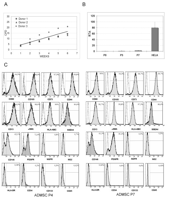
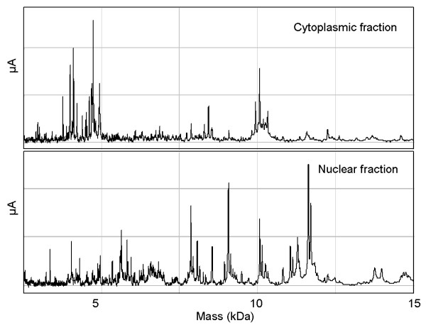
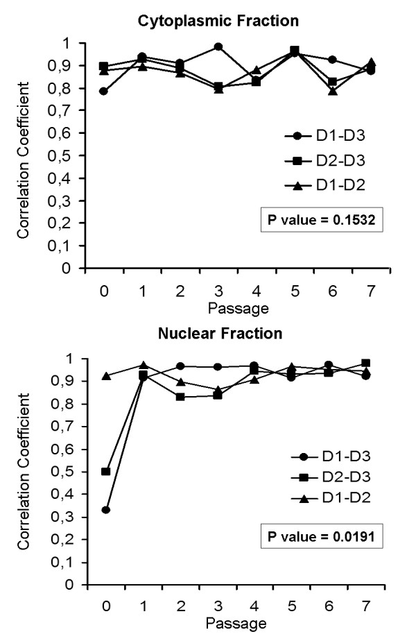
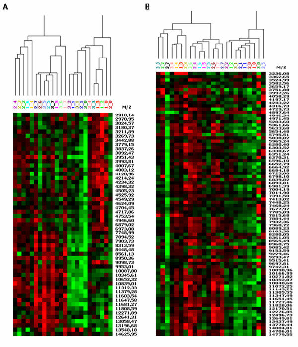
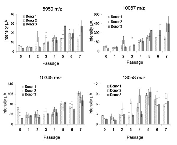
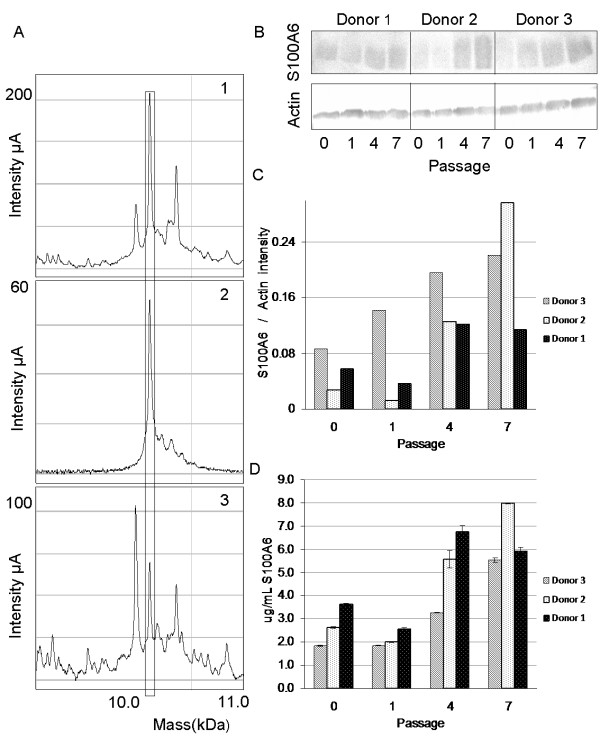
Results: We used Surface Enhanced Laser Desorption/Ionization-Time Of Flight-Mass Spectrometry (SELDI-ToF-MS) to evaluate the presence of stable molecular markers in adipose tissue-derived mesenchymal stem cells (AD-MSC) produced under conditions of good manufacturing practices (GMP). Proteomic patterns of cells prepared were consistent, with 4 up-regulated peaks (mass-to-charge ratio (m/z) 8950, 10087, 10345, and 13058) through subculture steps (P0-P7) with similar trend in three donors. Among the differentially expressed proteins found in the cytoplasmic and nuclear fractions, a cytoplasmic 10.1 kDa protein was upregulated during culture passages and was identified as S100A6 (Calcyclin).

Conclusions: This study suggests for the first time that common variation could occur in AD-MSC from different donors, with the identification of S100A6, a protein prevalently related to cell proliferation and cell culture condition. These results support the hypothesis of common proteomic changes during MSCs expansion and could give important insight in the knowledge of molecular mechanisms intervening during MSC expansion.


Figures

Figure 1
Figure 1
AD-MSC characterization. A) Cumulative population doublings of 3 AD-MSC lines. B) Telomerase activity in AD-MSC. The RTA was calculated from the different absorbances as follows: RTA = [(As sample-As heated-sample)]/[As Internal standard of the control template x 100]. It is representative of telomerase activity of 3 AD-MSC lines from different donors (median and range). C) Representative AD-MSC immunophenotype profile by flow cytometry at passages 4 and 7.
Figure 2
Figure 2
SELDI-ToF spectra of protein extract from different cellular compartments.
Figure 3
Figure 3
Donor-to-donor variability. Variation of the mean correlation coefficients (Pearson’s correlation) between each donor pair (D1: donor1, D2: donor2, D3: donor3) during subculturing passages (P0-P7) for cytoplasmic and nuclear enriched fraction.
Figure 4
Figure 4
Graphical representation of cluster analyses of SELDI-ToF mass spectrometry. Peaks detected in AD-MSC from different donors (1-2-3) during subculturing passages (from 0 to 7) indicated as donor_passage for A) cytoplasmic and B) nuclear enriched fraction. Red color intensity, high expressed; green color intensity, low expressed.
Figure 5
Figure 5
Protein peaks having a similar trend in all the three donors during AD-MSC expansion.
Figure 6
Figure 6
Validation of S100A6 in cytoplasmic extract of AD-MSC. A) SELDI-ToF immunoassay of m/z 10,087. Cytoplasmic extract from a pool of AD-MSC at passage 7: 1) spotted on CM10 array; 2) spotted on PS10 array indirectly coupled to anti-S100A6 antibody; 3) eluted from PS10 array indirectly coupled to anti-S100A6 antibody and spotted on CM10 array. The signals enclosed by the rectangle represent the peak at m/z 10,087. B) SDS-PAGE/Western blot analysis using anti-S100A6 and anti-Actin antibody of AD-MSC during different passages of cell expansion for different donors. C) Western blot densitometry quantification of the corresponding S100A6. Values are expressed as relative intensity ratio between S100A6 and Actin. D) Human S100-A6 ELISA quantification. Values are expressed as absolute quantification (μg/ml S100A6).

References

    1. Pittenger MF, Mackay AM, Beck SC, Jaiswal RK, Douglas R, Mosca JD, Moorman MA, Simonetti DW, Craig S, Marshak DR. Multilineage potential of adult human mesenchymal stem cells. Science. 1999;284:143–147. doi: 10.1126/science.284.5411.143. - DOI - PubMed
    1. Erices A, Conget P, Minguell JJ. Mesenchymal progenitor cells in human umbilical cord blood. Br J Haematol. 2000;109:235–242. doi: 10.1046/j.1365-2141.2000.01986.x. - DOI - PubMed
    1. Zuk PA, Zhu M, Ashjian P, De Ugarte DA, Huang JI, Mizuno H, Alfonso ZC, Fraser JK, Benhaim P, Hedrick MH. Human adipose tissue is a source of multipotent stem cells. Mol Biol Cell. 2002;13:4279–4295. doi: 10.1091/mbc.E02-02-0105. - DOI - PMC - PubMed
    1. Digirolamo CM, Stokes D, Colter D, Phinney DG, Class R, Prockop DJ. Propagation and senescence of human marrow stromal cells in culture: a simple colony-forming assay identifies samples with the greatest potential to propagate and differentiate. Br J Haematol. 1999;107:275–281. doi: 10.1046/j.1365-2141.1999.01715.x. - DOI - PubMed
    1. Gronthos S, Zannettino AC, Hay SJ, Shi S, Graves SE, Kortesidis A, Simmons PJ. Molecular and cellular characterisation of highly purified stromal stem cells derived from human bone marrow. J Cell Sci. 2003;116:1827–1835. doi: 10.1242/jcs.00369. - DOI - PubMed