. 2012 Oct;20(10):1893-901.
doi: 10.1038/mt.2012.118. Epub 2012 Jul 24.

Merits of combination cortical, subcortical, and cerebellar injections for the treatment of Niemann-Pick disease type A

Affiliations

Merits of combination cortical, subcortical, and cerebellar injections for the treatment of Niemann-Pick disease type A

Jie Bu et al. Mol Ther. 2012 Oct.

Abstract

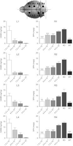
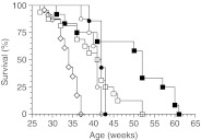
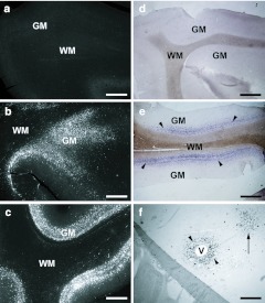
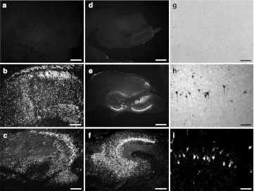
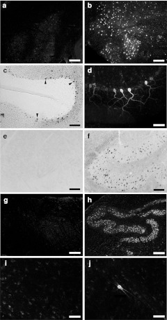
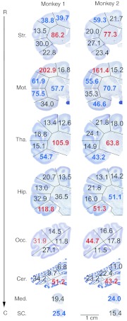
Niemann-Pick disease Type A (NPA) is a neuronopathic lysosomal storage disease (LSD) caused by the loss of acid sphingomyelinase (ASM). The goals of the current study are to ascertain the levels of human ASM that are efficacious in ASM knockout (ASMKO) mice, and determine whether these levels can be attained in non-human primates (NHPs) using a multiple parenchymal injection strategy. Intracranial injections of different doses of AAV1-hASM in ASMKO mice demonstrated that only a small amount of enzyme (<0.5 mg hASM/g tissue) was sufficient to increase survival, and that increasing the amount of hASM did not enhance this survival benefit until a new threshold level of >10 mg hASM/g tissue was reached. In monkeys, injection of 12 tracts of AAV1-hASM resulted in efficacious levels of enzyme in broad regions of the brain that was aided, in part, by axonal transport of adeno-associated virus (AAV) and movement through the perivascular space. This study demonstrates that a combination cortical, subcortical, and cerebellar injection protocol could provide therapeutic levels of hASM to regions of the NHP brain that are highly affected in NPA patients. The information from this study might help design new AAV-mediated enzyme replacement protocols for NPA and other neuronopathic LSDs in future clinical trials.


Figures

Figure 1
Figure 1
Levels of hASM enzyme and SPH lipid following intracranial injection of different doses of AAV1-hASM in ASMKO mice. Before homogenization, the right and left hemispheres were separated and further divided into four coronal slabs. Brain slabs that contained an injection site were marked with a black circle in the above diagram. ELISA assays (left column) showed a dose-dependent increase in hASM levels throughout the brain. SPH assays (right column) showed a significant reduction in SPH levels in all the brain slabs following treatment with the highest (1.2 × 10¹¹ gc) and both intermediate (4 × 10¹⁰, 1 × 10¹⁰ gc) doses compared to untreated ASMKO. Furthermore, mice administered the highest dose was the only group that contained SPH levels that was not statistically different than WT mice in all the brain slabs (P > 0.05), indicating that global reduction of SPH to near normal levels was achieved with 1.2 × 10¹¹ gc of AAV1-hASM. Although the lowest dose (4 × 10⁹ gc) provided a decrease in brain storage, the SPH levels were not significantly different from untreated ASMKO mice (P > 0.05). The statistical analysis used was a one-way variance (ANOVA) and Bonferroni multiple post hoc comparison (*P < 0.05; ** P < 0.01; ***P < 0.001), n = 6 for each group. AAV, adeno-associated virus; ANOVA, analysis of variance; ASMKO, acid sphingomyelinase knockout; ELISA, enzyme-linked immunosorbent assay; gc, genome copies; SPH, sphingomyelin; WT, wild-type.
Figure 2
Figure 2
All doses of AAV1-hASM improved the median survival of ASMKO mice. Shown is a Kaplan–Meier survival curve of untreated ASMKO mice (n = 23, open diamonds), and treated ASMKO mice that received a dose of 1.2 × 10¹¹ (n = 12, closed squares), 4.0 × 10¹⁰ (n = 16, open squares), 1.2 × 10¹⁰ (n = 7, closed circles), and 4.0 × 10⁹ (n = 8, open circles) genome copies per mouse. All treated ASMKO mice showed a significant improvement in median survival compared to ASMKO controls (P < 0.0001). AAV, adeno-associated virus; ASMKO, acid sphingomyelinase knockout.
Figure 3
Figure 3
Cortical injections of AAV1-hASM in NHPs. (ac) Immunohistochemistry and (df) in situ hybridization of the (a,b) motor cortex and the (ce) occipital cortex in (a,d) saline-treated and (b,c,e,f) AAV1-hASM–treated monkeys. The arrowheads in e point to a band of in situ hybridization-positive cells in the gray matter (purple color) immediately adjacent to the white matter. In situ hybridization in a region of the brain outside the injection sites showed hASM mRNA expression surrounding blood vessels (f, arrowheads). In some cases, a similar pattern of gene expression could also be observed outside the plane of the blood vessel (arrow). GM, gray matter; V, blood vessel; WM, white matter. Bars: 1.0 mm (in ac); 2.0 mm (in d,e); 0.5 mm (in f). AAV, adeno-associated virus; NHP, non-human primate.
Figure 4
Figure 4
Subcortical injection of AAV1-hASM in NHPs. (af,i) Immunohistochemistry and (g,h) in situ hybridization of the (a,b) striatum, (c) thalamus, (df) hippocampus, and (gi) entorhinal cortex in (a,d,g) saline-treated and (b,c,e,f,h,i) AAV1-hASM–treated monkeys. Bars: 1.0 mm (in ac); 2.0 mm (in d,e); 0.5 mm (in f); 0.2 mm (in gi). AAV, adeno-associated virus; NHP, non-human primate.
Figure 5
Figure 5
Cerebellar injection of AAV1-hASM in NHPs. (a,b,d,gj) Immunohistochemistry and (c,e,f) in situ hybridization of the (a,b) DCN injection site, (c,d) the cerebellar cortex, (eh) the medulla oblongata, and (i,j) the spinal cord. (c) The arrowheads point to examples of transduced cells (d) that contained classical Purkinje cell morphology in the correct laminar layer. (f,h) Clear evidence of retrograde axonal transport of AAV followed by hASM expression was observed in the medulla oblongata. (j) Human ASM protein was also observed in large neurons of the spinal cord. Bars: 0.2 mm (in ac,e,f); 0.05 mm (in d,i,j); 0.4 mm (in g,h). AAV, adeno-associated virus; DCN, deep cerebellar nucleus; NHP, non-human primate.
Figure 6
Figure 6
Summary of the total ASM levels along the rostral (R) to caudal (C) axis of the cynomolgus brain, as determined by ELISA assay. The baseline level in the saline-treated brain was 23.745 ng/mg ± 6.853 ng ASM/mg tissue (mean ± SD). Shown are the total ASM values (monkey ASM + AAV1 vector-derived hASM) in different brain slabs at the level of the injected striatum (Str.), motor cortex (Mot.), thalamus (Tha.), hippocampus (Hip.), occipital cortex (Occ.), and cerebellum (Cer.). Total ASM levels in brain slabs that contained an injection site are shown in red, which ranged from 4.0–26.1 (monkey 1) and 2.8–20.1 (monkey 2) SDs above the baseline mean. The only exception was the injected occipital cortex of monkey 1, which did not result in a significant increase in total ASM for unknown reasons. Brain slabs outside the injection sites that contained total ASM levels that were at least 2.0 SDs above the baseline mean are shown in blue. Brain slabs that were below 2.0 SDs were not significantly elevated and are shown in black. The medulla oblongata (Med.) and spinal cord (SC.) are contiguous structures that contained a low baseline level of 12.340 ± 4.904 ng ASM/mg tissue (mean ± SD) in the saline-treated monkey. DCN injections with AAV1-hASM resulted in total ASM levels that were 2.4 and 2.7 SDs above the baseline mean in the medulla oblongata of monkey 2 and the spinal cord of monkey 1, respectively. AAV, adeno-associated virus; ASM, acid sphingomyelinase; DCN, deep cerebellar nucleus.

References

    1. Schultz ML, Tecedor L, Chang M., and, Davidson BL. Clarifying lysosomal storage diseases. Trends Neurosci. 2011;34:401–410. - PMC - PubMed
    1. Sands MS., and, Haskins ME. CNS-directed gene therapy for lysosomal storage diseases. Acta Paediatr Suppl. 2008;97:22–27. - PMC - PubMed
    1. Sands MS., and, Davidson BL. Gene therapy for lysosomal storage diseases. Mol Ther. 2006;13:839–849. - PubMed
    1. Gray SJ, Matagne V, Bachaboina L, Yadav S, Ojeda SR., and, Samulski RJ. Preclinical differences of intravascular AAV9 delivery to neurons and glia: a comparative study of adult mice and nonhuman primates. Mol Ther. 2011;19:1058–1069. - PMC - PubMed
    1. Janson C, McPhee S, Bilaniuk L, Haselgrove J, Testaiuti M, Freese A.et al. (2002Clinical protocol: gene therapy of Canavan disease: AAV-2 vector for neurosurgical delivery of aspartoacylase gene (ASPA) to the human brain Hum Gene Ther 131391–1412. - PubMed

Substances