. 2012 Aug 24;425(2):189-94.
doi: 10.1016/j.bbrc.2012.07.064. Epub 2012 Jul 22.

Generation and characterization of Tmeff2 mutant mice

Affiliations

Generation and characterization of Tmeff2 mutant mice

Tian Rui Chen et al. Biochem Biophys Res Commun. .

Abstract

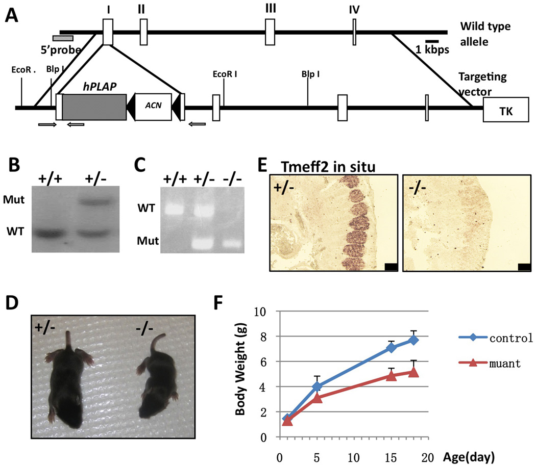
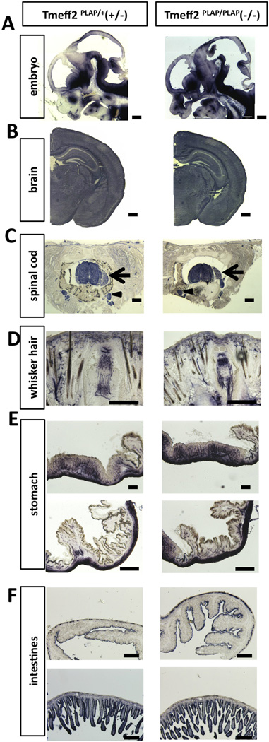
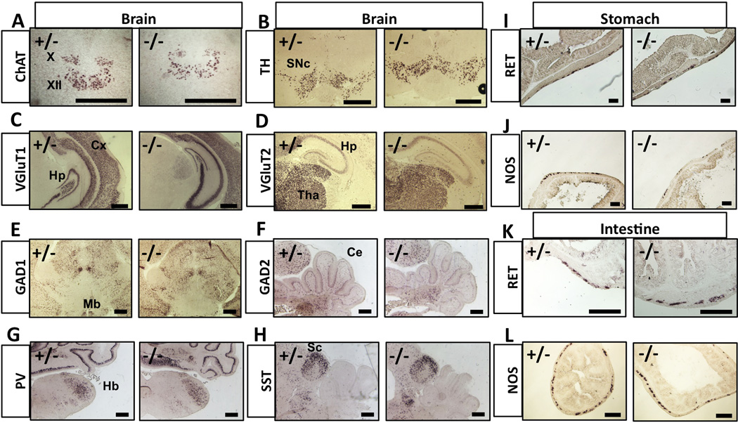
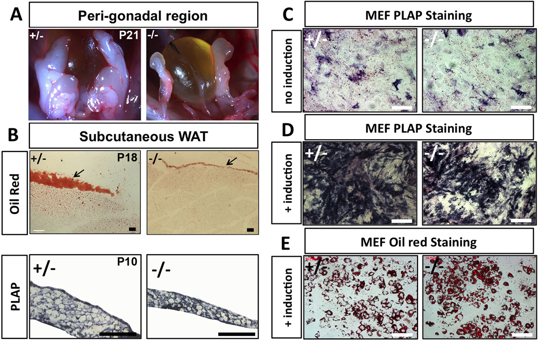
TMEFF2 is a single-transmembrane protein containing one EGF-like and two follistatin-like domains. Some studies implicated TMEFF2 as a tumor suppressor for prostate and other cancers, whereas others reported TMEFF2 functioning as a growth factor for neurons and other cells. To gain insights into the apparently conflicting roles of TMEFF2, we generated a null allele of Tmeff2 gene by replacing its first coding exon with human placental alkaline phosphatase cDNA (Tmeff2(PLAP)). Tmeff2(PLAP/PLAP) homozygous mutant mice are born normal, but show growth retardation and die around weaning age. Tmeff2 is widely expressed in the nervous system, and the Tmeff2(PLAP) knock-in allele enables the visualization of neuronal innervations of skin and internal organs with a simple alkaline phosphatase staining. Tmeff2 is also highly expressed in prostate gland and white adipose tissues (WAT). However, with the exception of reduced WAT mass, extensive anatomical and molecular analyses failed to detect any structural or molecular abnormalities in the brain, the spinal cord, the enteric nervous system, or the prostate in the Tmeff2 mutants. No tumors were found in Tmeff2-mutant mice. The Tmeff2(PLAP/PLAP) knock-in mouse is an useful tool for studying the in vivo biological functions of TMEFF2.


Figures

Figure 1
Figure 1. Generating Tmeff2PLAP knock-in allele and Tmeff2-KO mice
A. Schematic representation of the targeting vector and strategy. cDNA encoding hPLAP together with the ACN (neo) cassette were used to replace start codon ATG and the rest of exon 1 of the Tmeff2 gene. Exons are represented as white boxes. The negative selection thymidine kinase cassette is designated as TK. Arrows indicate the position of primers used for PCR genotyping analysis. B. Southern blot analysis of the genomic DNA from wildtype and targeted embryonic stem cells. C. PCR analysis of the genotypes. D. Compared to heterozygous littermate, Tmeff2-KO mice are smaller in size. E. Representative images of Tmeff2 in situ hybridization results showing the expression of Tmeff2 in dorsal root ganglion (DRG) in control embryo, and the lack of in situ signal in mutant embryo. Scale bar: 100µm. F. The growth curve of Tmeff2PLAP/+ (control) and Tmeff2-KO (mutant) mice (averaged from n>4 for each genotype at each age).
Figure 2
Figure 2. Tmeff2-KO mice have structurally normal central, peripheral and enteric nervous system as revealed by AP-staining
A. Representative sagittal sections of the head from Tmeff2PLAP/+ (+/−) and Tmeff2-KO (−/−) embryos (E12.5) stained for PLAP activity. B. Representative coronal sections of brains from P15 control (+/−) and Tmeff2-KO (−/−) mice stained for PLAP activity showing similar staining patterns. C. Representative coronal sections of spinal cord from control (+/−) and Tmeff2-KO (−/−) mice showing Tmeff2 expression in spinal cord, dorsal root ganglion (arrows), and sympathetic ganglion (arrowheads). No difference in staining patterns was observed between control and mutant mice. D. Representative coronal sections of whiskers from control (+/−) and Tmeff2-KO (−/−) mice stained for PLAP activity showing similar innervation patterns. E. Representative images of PLAP-stained stomach sections from heterozygous control (+/−) and Tmeff2-KO (−/−) mice. F. Representative images of PLAP-stained intestinal sections from control (+/−) and Tmeff2-KO (−/−) mice. Upper panels, large intestines; lower panels, small intestines. Scale bars: A, B 200µm. C, D, E, F 100µm.
Figure 3
Figure 3. Neuronal differentiations are grossly normal in Tmeff2-KO mice
A–H. Representative brain images of in situ hybridization results using probes against ChAT, TH, VGluT1, VGluT2, GAD1, GAD2, Parv or SST are shown here. X, vagus nuclei; XII, hypoglossal nuclei; SNc, substantia nigra; Cx, cortex; Hp, hippocampus; Tha, thalamus; Mb, midbrain; Ce, cerebellum; Hb, hindbrain; Sc, superior colliculus. I–J. Representative images of RET (I) or Nos1 (J) in situ hybridization results on sections of stomachs from control (+/−) and Tmeff2-KO (−/−) mice. K–L. Representative images of RET (K) or Nos1 (L) in situ hybridization results on sections of intestines from control (+/−) and Tmeff2-KO (−/−) mice. Scale bars: 100µm.
Figure 4
Figure 4. Tmeff2-KO mice have reduced WAT mass but TMEFF2 deficiency does not affect in vitro adipocyte differentiation
A. Representative images showing peri-gonadal white adipose tissues (WAT) in control heterozygous (+/−) and Tmeff2-KO (−/−) mice. Tmeff2-KO mice contain almost no visible WAT at the age of P21 (dying animals). B. Sections of subcutaneous WAT and neighboring tissues stained with Oil Red showing residual amount of WAT in Tmeff2-KO animals at age P18 (upper panels). AP-staining on sections of dissected WAT from control (+/−) and Tmeff2-KO (−/−) animals (age=P10) showing Tmeff2 expression in adipocytes (lower panels). Scale bars: 100µm. C. Low level spontaneous expression of Tmeff2 is revealed by PLAP staining during regular cultures of MEFs isolated from heterozygous (+/−) and Tmeff2-KO (−/−) mice. D. During in vitro induction of MEFs differentiation into adipocytes, Tmeff2 expression is drastically increased as revealed by PLAP staining. E. Tmeff2-KO (−/−) MEFs are equally competent as heterozygous (+/−) MEFs to differentiate into adipocytes upon induction as revealed by Oil Red staining.

Similar articles

Cited by

References

    1. Uchida T, Wada K, Akamatsu T, Yonezawa M, Noguchi H, Mizoguchi A, Kasuga M, Sakamoto C. A novel epidermal growth factor-like molecule containing two follistatin modules stimulates tyrosine phosphorylation of erbB-4 in MKN28 gastric cancer cells. Biochem Biophys Res Commun. 1999;266:593–602. - PubMed
    1. Liang G, Robertson KD, Talmadge C, Sumegi J, Jones PA. The gene for a novel transmembrane protein containing epidermal growth factor and follistatin domains is frequently hypermethylated in human tumor cells. Cancer research. 2000;60:4907–4912. - PubMed
    1. Horie M, Mitsumoto Y, Kyushiki H, Kanemoto N, Watanabe A, Taniguchi Y, Nishino N, Okamoto T, Kondo M, Mori T, Noguchi K, Nakamura Y, Takahashi E, Tanigami A. Identification and characterization of TMEFF2, a novel survival factor for hippocampal and mesencephalic neurons. Genomics. 2000;67:146–152. - PubMed
    1. Young J, Biden KG, Simms LA, Huggard P, Karamatic R, Eyre HJ, Sutherland GR, Herath N, Barker M, Anderson GJ, Fitzpatrick DR, Ramm GA, Jass JR, Leggett BA. HPP1: a transmembrane protein-encoding gene commonly methylated in colorectal polyps and cancers. Proceedings of the National Academy of Sciences of the United States of America. 2001;98:265–270. - PMC - PubMed
    1. Glynne-Jones E, Harper ME, Seery LT, James R, Anglin I, Morgan HE, Taylor KM, Gee JM, Nicholson RI. TENB2, a proteoglycan identified in prostate cancer that is associated with disease progression and androgen independence, International journal of cancer. Journal international du cancer. 2001;94:178–184. - PubMed

Publication types

LinkOut - more resources