Review
. 2012 Jul 25;12(8):570-80.
doi: 10.1038/nri3261.

Lighting the fires within: the cell biology of autoinflammatory diseases

Affiliations
Review

Lighting the fires within: the cell biology of autoinflammatory diseases

Heiyoung Park et al. Nat Rev Immunol. .

Abstract

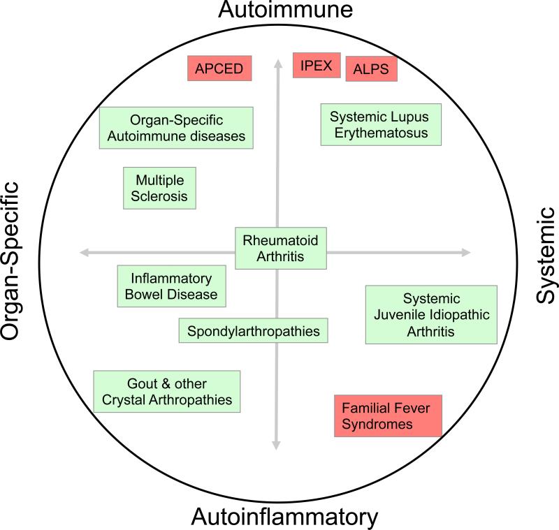
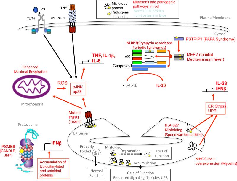
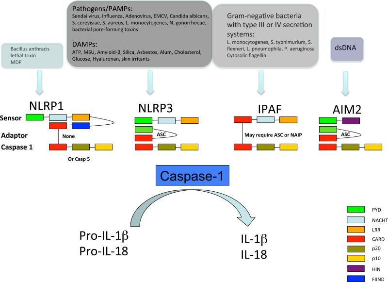
Autoinflammatory diseases are characterized by seemingly unprovoked pathological activation of the innate immune system in the absence of autoantibodies or autoreactive T cells. Discovery of the causative mutations underlying several monogenic autoinflammatory diseases has identified key regulators of innate immune responses. Recent studies have highlighted the role of misfolding, oligomerization and abnormal trafficking of pathogenic mutant proteins in triggering autoinflammation, and suggest that more common rheumatic diseases may have an autoinflammatory component. This coincides with recent discoveries of new links between endoplasmic reticulum stress and inflammatory signalling pathways, which support the emerging view that autoinflammatory diseases may be due to pathological dysregulation of stress-sensing pathways that normally function in host defence.


Figures

Figure 1
Figure 1. The spectrum of autoimmune and autoinflammatory diseases
Polygeneic diseases are boxed in green and menedelian disorders boxed in red. The horizontal axis depicts the range of organ-specfic vs. systemic disease, and the vertical axis, the degree of involvement of the parts of the immune system in immunopathology: innate (autoinflammatory, bottom) vs. adaptive (autoimmune, top).
Figure 2
Figure 2. Consequences of protein misfolding and intracellular signaling complexes that activate autoinflammatory disease
A) The outcomes of misfolding of secretory protewins in the ER are depicted at the bottom of the figure. Degradation of misfolded protein can cause a loss of function, whereas accumulation of misfolded proteins can trigger abnormal intracellular signaling, or at higher levels, induction of the unfolded protein response (UPR), which can also lead to the induction of inflammation and programmed cell death. Different foci of abnormal cellular signaling that trigger autoinflammatory diseases are depicted in the cell, clockwise from lower left. B) In PSMB8 deficiency, reduced degradation of misfolded proteins and peptides by the immunoproteasome leads to accumulation of ubiquinated proteins and cellular stress, which leads of Interferon-β production. Type I Interferon in turn upregulates the synthesis of immunoproteasome sububits, perpetuating the abnormalities. C) In TRAPS, extracellular mutations in TNFR1 lead to accumulation of the mutant receptor in the ER, which triggers an abnormal inflammatory response that is amplified by TNF or LPS signaling through cell surface receptors. D) In the Cryopyrin-Associated Perodic Syndromes (CAPS), mutations in NLRP3 enhance activation of the NLRP3 inflammasome and processing of IL-1β into its active form. E) In the spondylarthropathies, high-level expression of HLA-B27, which is enhanced in inflammation, fails to fold properly and is retained in the ER, triggering a partial ER stress response which leads to type I interferon and IL-23 production.
Figure 3
Figure 3. The NLRP1, NLRP3, IPAF and AIM2 inflammasomes
The NLRP1 inflammasome is activated by Bacillus anthracis lethal toxin and muramyl dipeptide (MDP). The NLRP3 inflammasome is activated by exposure to whole pathogens, pathogen-associated molecular patterns (PAMPs), damage-associated molecular patterns (DAMPs) and environmental irritants. The IPAF inflammasome is activated by Gram-negative bacteria with type III or IV secretion systems, and the DNA inflammasome senses double stranded DNA. Other complexes, such as NLRP6-containing complexes that regulate IL-18, also exist , and pyrin likely regulates an inflammasome complex containing ASC but not NLRP3 that activates IL-1β processing . EMCV – Encephalomyocarditis virus, ATP – Adenosine triphosphate, MSU – Monosodium urate.

References

    1. McGonagle D, McDermott MF. A proposed classification of the immunological diseases. PLoS Med. 2006;3:e297. - PMC - PubMed
    1. Masters SL, Simon A, Aksentijevich I, Kastner DL. Horror autoinflammaticus: the molecular pathophysiology of autoinflammatory disease. Annu Rev Immunol. 2009;27:621–668. - PMC - PubMed
    1. Martinon F, Petrilli V, Mayor A, Tardivel A, Tschopp J. Gout-associated uric acid crystals activate the NALP3 inflammasome. Nature. 2006;440:237–241. [This paper connected the pathogenesis of gout to activation of the NLRP3 inflammasome by uric acid crystals] - PubMed
    1. Schroder M, Kaufman RJ. ER stress and the unfolded protein response. Mutat Res. 2005;569:29–63. - PubMed
    1. Pipe SW, Kaufman RJ. Factor VIII C2 domain missense mutations exhibit defective trafficking of biologically functional proteins. J Biol Chem. 1996;271:25671–25676. - PubMed

Publication types

MeSH terms