. 2011 Nov;1(11):e38.
doi: 10.1038/bcj.2011.37. Epub 2011 Nov 4.

Resistance of MLL-AFF1-positive acute lymphoblastic leukemia to tumor necrosis factor-alpha is mediated by S100A6 upregulation

Resistance of MLL-AFF1-positive acute lymphoblastic leukemia to tumor necrosis factor-alpha is mediated by S100A6 upregulation

H Tamai et al. Blood Cancer J. 2011 Nov.

Abstract

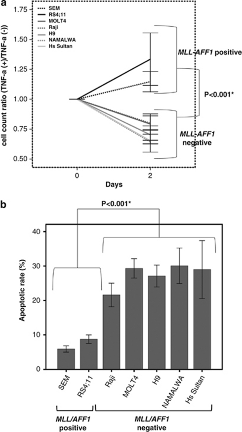
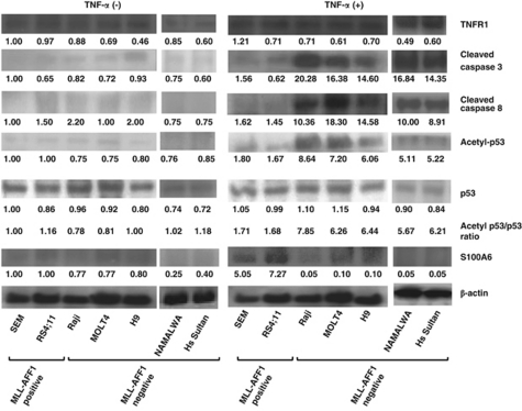
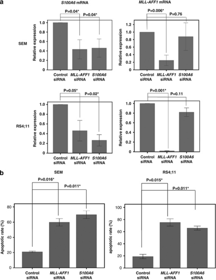
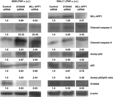
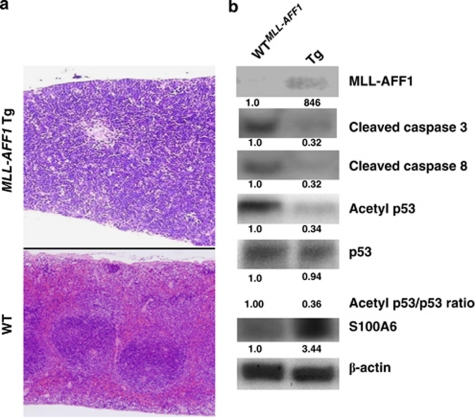
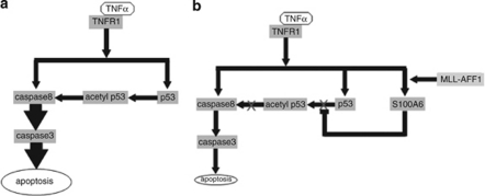
Mixed-lineage leukemia (MLL)-AFF1 (MLL-AF4)-positive acute lymphoblastic leukemia (ALL) is associated with poor prognosis, even after allogeneic hematopoietic stem cell transplantation (allo-HSCT). The resistance to graft-versus-leukemia (GVL) effects may be responsible for the poor effect of allo-HSCT on MLL-AFF1-positive ALL. Cytotoxic effector mechanisms mediated by tumor necrosis factor-alpha (TNF-α) was reported to contribute to the GVL effect. We showed that MLL-AFF1-positive ALL cell lines are resistant to TNF-α. To examine the mechanism of resistance to TNF-α of MLL-AFF1-positive leukemia, we focused on S100A6 as a possible factor. Upregulation of S100A6 expression and inhibition of the p53-caspase 8-caspase 3 pathway were observed only in MLL-AFF1-positive ALL cell lines in the presence of TNF-α. The effect of S100A6 on resistance to TNF-α by inhibition of the p53-caspase 8-caspase 3 pathway of MLL-AFF1-positive ALL cell lines were also confirmed by analysis using small interfering RNA against S100A6. This pathway was also confirmed in previously established MLL-AFF1 transgenic mice. These results suggest that MLL-AFF1-positive ALL escapes from TNF-α-mediated apoptosis by upregulation of S100A6 expression, followed by interfering with p53-caspase 8-caspase 3 pathway. These results suggest that S100A6 may be a promising therapeutic target for MLL-AFF1-positive ALL in combination with allo-HSCT.


Figures

Figure 1
Figure 1
The comparison of sensitivity to TNF-α between MLL–AFF1-positive ALL cell lines and MLL–AFF1-negative ALL cell lines. (a) Cell count ratio (TNF-α(+)/TNF-α(−)) of MLL–AFF1-negative cell line (SEM and RS4;11) and MLL–AFF1-negative cell line (MOLT4, Raji, H9, NAMALWA, Hs Sultan). TNF-α (5 ng/ml) significantly inhibited the proliferation of cell lines only in MLL–AFF1-negative cell lines (P<0.001). (b) Inhibition rate by TNF-α (5 ng/ml) of leukemia cell line. The apoptotic rate of MLL–AFF1-positive ALL cell lines 48 h after addition of TNF-α (5 ng/ml) were significantly lower than those of MLL–AFF1-negative leukemia cell lines.
Figure 2
Figure 2
Western blotting analysis of lysate from leukemia cell lines. (After 48 h of addition or no addition of TNF-α (5 ng/ml)). All the band were quantified and normalized by β-actin. Normalized densities of SEM cells without TNF-α were standardized as 1.00. Upregulation of S100A6 expression and downregulation of acetyl-p53/p53 expression ratio, cleaved caspase 3 and cleaved caspase 8 expression only in MLL–AFF1-positive ALL cell lines in the presence of TNF-α (5 ng/ml), although there were no significant differences between the two groups in the absence of TNF-α. There were no differences of TNF receptor 1 (TNFR1) expression in the absence or presence of TNF-α. Densitometry measurements were standardized by those of SEM without TNF-α.
Figure 3
Figure 3
Effect of S100A6 siRNA or MLL–AFF1 siRNA on MLL–AFF1-positive cell lines. (a) Real-time quantitative PCR analysis of S100A6 mRNA and MLL–AFF1 mRNA under S100A6 siRNA or MLL–AFF1 siRNA or control siRNA in the presence of TNF-α (5 ng/ml) of MLL–AFF1-positive cell lines (SEM and RS4;11). S100A6 mRNA expression was significantly inhibited by both MLL–AFF1 siRNA and S100A6 siRNA in comparison with control siRNA. MLL–AFF1 mRNA expression was significantly inhibited by MLL–AFF1 siRNA in comparison with control siRNA, but MLL–AFF1 expression was not significantly inhibited by S100A6 siRNA. (b) Apoptotic rate by S100A6 siRNA or MLL–AFF1 siRNA or control siRNA in the presence of TNF-α (5 ng/ml) of MLL–AFF1-positive cell lines (SEM and RS4;11). The apoptotic rate of MLL–AFF1-positive ALL cell lines treated with siRNA against MLL–AFF1 in the presence of TNF-α (5 ng/ml) was significantly higher than those treated with control siRNA. The apoptotic rate of MLL–AFF1-positive ALL cell lines treated with siRNA against S100A6 in the presence of TNF-α was significantly higher than those treated with mismatch control siRNA.
Figure 4
Figure 4
Western blotting analysis of lysate from MLL–AFF1-positive leukemia cell lines (SEM and RS4;11) under the MLL–AFF1 siRNA/S100A6 siRNA/control siRNA in the presence of TNF-α (5 ng/ml). All of acetyl-p53/p53 ratio, cleaved caspase 8 and cleaved caspase 3 expression were increased in cells treated with MLL–AFF1 siRNA or s100A6 siRNA in comparison with those treated with control siRNA in the presence of TNF-α (5 ng/ml). S100A6 expression was inhibited by both MLL–AFF1 siRNA and S100A6 siRNA in comparison with control siRNA. MLL–AFF1 expression was inhibited by MLL–AFF1 siRNA in comparison with control siRNA, but MLL–AFF1 expression was not inhibited by S100A6 siRNA. Densitometry measurements of control siRNA were standardized as 1.00.
Figure 5
Figure 5
Examination of S100A6 expression in MLL–AFF1 transgenic (Tg) mouse. (a) Histopathological findings from 14-month-old wild-type (WT) and MLL–AFF1 Tg mice (original magnification × 4). Comparison of their spleens shows infiltration of lymphoma cells and destruction of the normal organ structure in the MLL–AFF1 Tg mouse. (b)Western blotting analysis of lysate from spleen of WT or MLL–AFF1 Tg mice. Upregulation of S100A6 and inhibition of upregulation of the p53–caspase 8–caspase 3 pathway were also confirmed in this mouse model. Densitometry measurements were standardized by those of WT.
Figure 6
Figure 6
Working hypothesis of the effect of TNF-α on MLL–AFF1-positive ALL cell lines and those on MLL–AFF1-negative ALL cell lines. (a) Working hypothesis of the effect of TNF-α on MLL–AFF1-negative cell lines. TNF-α seems to lead leukemia cells to apoptosis through caspase 8–caspase 3 pathway or p53–caspase 8–caspase 3 pathway. (b) Working hypothesis of the pathway of MLL–AFF1-positive cell lines to be resistant to TNF-α. MLL–AFF1-positive ALL cell lines seem to be resistant to TNF-α by upregulation of S100A6 via inhibition of upregulation of the p53–caspase 8–caspase 3 pathway.

Similar articles

Cited by

References

    1. Harper DP, Aplan PD. Chromosomal rearrangements leading to MLL gene fusions: clinical and biological aspects. Cancer Res. 2008;68:10024–10027. - PMC - PubMed
    1. Pui CH, Gaynon PS, Boyett JM, Chessells JM, Baruchel A, Kamps W, et al. Outcome of treatment in childhood acute lymphoblastic leukaemia with rearrangements of the 11q23 chromosomal region. Lancet. 2002;359:1909–1915. - PubMed
    1. Ringdén O, Karlsson H, Olsson R, Omazic B, Uhlin M. The allogeneic graft-versus-cancer effect. Br J Haematol. 2009;147:614–633. - PubMed
    1. Sastry M, Ketchem RR, Crescenzi O, Weber C, Lubienski MJ, Hidaka H. The three-dimensional structure of Ca(2+)-bound calcyclin: implications for Ca(2+)-signal transduction by S100 proteins. Structure. 1998;6:223–231. - PubMed
    1. Tsoporis JN, Izhar S, Parker TG. Expression of S100A6 in cardiac myocytes limits apoptosis induced by tumor necrosis factor-alpha. J Biol Chem. 2008;283:30174–30183. - PMC - PubMed