. 2012 Jul;24(7):2839-56.
doi: 10.1105/tpc.112.101022. Epub 2012 Jul 24.

Nuclear ribosome biogenesis mediated by the DIM1A rRNA dimethylase is required for organized root growth and epidermal patterning in Arabidopsis

Affiliations

Nuclear ribosome biogenesis mediated by the DIM1A rRNA dimethylase is required for organized root growth and epidermal patterning in Arabidopsis

Yana Wieckowski et al. Plant Cell. 2012 Jul.

Abstract

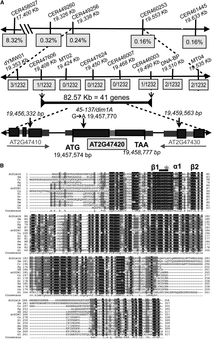
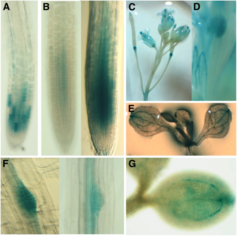
Position-dependent patterning of hair and non-hair cells in the Arabidopsis thaliana root epidermis is a powerful system to study the molecular basis of cell fate specification. Here, we report an epidermal patterning mutant affecting the ADENOSINE DIMETHYL TRANSFERASE 1A (DIM1A) rRNA dimethylase gene, predicted to participate in rRNA posttranscriptional processing and base modification. Consistent with a role in ribosome biogenesis, DIM1A is preferentially expressed in regions of rapid growth, and its product is nuclear localized with nucleolus enrichment. Furthermore, DIM1A preferentially accumulates in the developing hair cells, and the dim1A point mutant alters the cell-specific expression of the transcriptional regulators GLABRA2, CAPRICE, and WEREWOLF. Together, these findings suggest that establishment of cell-specific gene expression during root epidermis development is dependent upon proper ribosome biogenesis, possibly due to the sensitivity of the cell fate decision to relatively small differences in gene regulatory activities. Consistent with its effect on the predicted S-adenosyl-l-Met binding site, dim1A plants lack the two 18S rRNA base modifications but exhibit normal pre-rRNA processing. In addition to root epidermal defects, the dim1A mutant exhibits abnormal root meristem division, leaf development, and trichome branching. Together, these findings provide new insights into the importance of rRNA base modifications and translation regulation for plant growth and development.


Figures

Figure 1.
Figure 1.
Cell-Type Expression of GL2pro:GUS Is Altered in 45-137/dim1A. (A) Diagram of a transverse section of an Arabidopsis root meristem. The primary root is organized in concentric rings of cells including the endodermis (En), cortex (C), and the epidermis (which is comprised of non hair cells [N] and hair cells [H]). (B) Current model of epidermal patterning in Arabidopsis. Note: solid lines represent gene regulation, and dashed lines indicate protein movement. (C) Analysis of position-dependent expression of GL2 using promoter-reporter transgene GL2pro:GUS in the wild type (WT; Ws), the 45-137/dim1A mutant, two dim1A complementation lines, two DIM1Apro:DIM1A-GFP lines, and the wild type treated with 5 µg/mL cycloheximide. Images are representative roots showing the expression pattern of GL2pro:GUS in the root meristem of 4-d-old seedlings. (D) Quantification of the epidermal cell-type pattern, showing frequencies of ectopic GL2pro:GUS-expressing cells in the H cell position (gray bars) and ectopic non-GL2pro:GUS–expressing cells in the N cell position (black bars), for each line. The mean and sd are indicated for each line. Complementation lines A- and B- are included as negative controls for complementation lines A and B, respectively. Symbols indicate a value significantly different than the corresponding wild-type (WT) value (using t test): asterisk, P < 0.01.
Figure 2.
Figure 2.
Mapping and Identification of a Mutant of the Arabidopsis DIM1A Gene. (A) Fine mapping using individual F2 plants delimited the 45-137 mutation to an 83-kb region on the lower arm of chromosome 2 between markers CER447606 and CER446003. At2g47420 contains two exons (black bars) and one intron (gray bar), and it encodes DIM1A, a putative rRNA dimethyladenosine transferase. (B) Multiple alignment of the amino acid sequences from eukaryotic DIM1A homologs. The highly conserved β sheets (β1 and β2) and α helix (α1) important for SAM binding are indicated. Binding “motif 1” (shown as a black bar) contains the canonical GXGXG SAM binding sequence. The asterisk within motif 1 indicates the position of the mutation in dim1A. Arabidopsis (At), Zea mays (Zm), Chlamydomonas reinhardtii (Cr), Physcomitrella patens (Pp), Dictyostelium dictoideim (Dd), S. cerevisiae (Sc), Cryptococcus neoformans (Cn), Xenopus laevis (Xl), Danio rerio (Dr), Homo sapiens (Hs), Drosophila melanogaster (Dm), Caenorhabditus elegans (Ce), and Giardia lambia (Gl). Black shading indicates 90% consensus, gray shading indicates 50% consensus, the exclamation point is any one of IV, the dollar sign is any one of LM, the percent symbol is any one of FY, and the hash tag is any one of NDQEBZ.
Figure 3.
Figure 3.
The dim1A Mutant Lacks Two Conserved 18S rRNA Adenosine Dimethylations. (A) Predicted sequence and structure of the 3′-end of the Arabidopsis 18S rRNA. The arrow represents the reverse complement of the 16-bp primer used for poisoned primer extension. (B) Poisoned primer extension products using RNA isolated from the wild type and dim1A. The left side of the gel shows the RNA sequencing reaction for reference. Primer extension yielded a signal at the adenosine doublet in the wild type (WT; indicated by the arrowhead). In the dim1A mutant, there is no indication of a reverse transcriptase stop at either of the adenosines, but rather termination is seen at the next uridine in the 18S rRNA sequence (product indicated by arrow). In both (A) and (B), “A” represent the adenosines that are putatively dimethylated and U* indicates the uridine at which the reaction was forced to stop due to the presence of ddATP in place of dATP in the nucleotide mixture.
Figure 4.
Figure 4.
The dim1A Mutation Does Not Affect 35S pre-rRNA Processing. (A) Structure of Arabidopsis pre-rRNA transcript and predicted processing sites. The 35S pre-rRNA is composed of the 5′ ETS, 18S rRNA (18S), ITS1, 5.8S rRNA (5.8S), ITS2, 25S rRNA (25S), and 3′ ETS. The primers U1 and U2 were designed to detect processing at site P, primers U2 and U3 to detect the 5′ ETS, and primers IS1 and IS2 to detect ITS1. (B) Total RNA was purified from 5-d-old wild-type and dim1A plants and subjected to RT-PCR using primers specific for 5′ ETS, ITS1, 18S, and translation elongation factor EF1αA4 (EF14a) as a control. Based on knowledge of Dim1-dependent processing in yeast, DIM1A putatively processes the 35S pre-rRNA at sites A1 and A2, which can be monitored using primer sets U2+U3 and IS1+IS2, respectively. No significant difference in product accumulation between the wild type (WT) and dim1A was observed at 24, 28, or 32 PCR cycles, suggesting that the dim1A mutation does not alter the pre-rRNA processing activity of the DIM1A enzyme.
Figure 5.
Figure 5.
DIM1A Is Expressed in Developing Tissues. Histochemical GUS staining pattern of transgenic plants carrying the DIM1Apro:GUS transgene. T3 plants identified as homozygous for DIM1Apro:GUS were observed: (A) 2-d-old root tip; (B) 4-d-old root tips stained for moderate (left) and extended (right) times; (C) floral tissue (stigma and petals); (D) anther, pollen grains, and sepal vasculature; (E) developing leaves and cotyledon of 10-d-old plant; (F) lateral root emergence site and vascular tissue staining in two different 4-d-old seedlings; and (G) 2-d-old cotyledon.
Figure 6.
Figure 6.
DIM1Apro:DIM1A-GFP Localizes to the Nucleus and Nucleolus of Root Cells. Subcellular and root tissue localization of DIM1A-GFP expressed from the DIM1A promoter of 4-d-old seedlings. Seedlings were stained with PI (red) for visualizing the cell wall and DIM1Apro:DIM1A-GFP accumulation (green). Bars = 20 μm. (A) and (B) Root meristem localization of free GFP (A) and DIM1Apro:DIM1A-GFP (B). (C) Overlay of fluorescence image and differential interference contrast (DIC) optics. (D) to (F) DIM1Apro:DIM1A-GFP persists longer in developing hair cells (asterisks) than non-hair cells ([D] and [E]) and is present in all meristematic tissues (F). C, cortex; En, endodermis; Ep, epidermis; S, stele.
Figure 7.
Figure 7.
Genetic Interaction of dim1A and Epidermal Patterning Genes. (A) to (D) Expression pattern of the following transcription factor reporter genes in roots of wild-type (WT) and dim1A 4-d-old seedlings: CPCpro:GUS (A), EGL3pro:GUS (B), and WERpro:GFP ([C] and [D]). Images in (D) represent magnification of region within boxes diagrammed in (C). Bars = 50 μm in (A), (B), and (D) and 25 μm in (C). (E) Graphical representation of the ratio of the amount of GFP (expressed from the WER promoter) measured in cells in the N position to the amount measured in cells in the H position in wild-type (gray bars) and dim1A roots (black bars). GFP pixel quantification was done in cells in the uppermost (shootward) one-third of the meristem. Measurements are expressed in terms of total pixels (left) and pixels/area (right). The mean and sd are indicated for each line. (F) and (G) The genetic interaction of dim1A and epidermal patterning mutants using expression level and pattern of GL2pro:GUS as a readout. (F) Expression of the GL2pro:GUS transcriptional reporter in the following lines: the wild type and dim1A; ttg1 and dim1A ttg1; gl3 egl3 and dim1A gl3 egl3; wer and dim1A wer; scm-2 and dim1A scm-2; and gl2 and dim1A gl2. Bars = 50 μm. (G) Quantification of the epidermal cell-type pattern, showing frequencies of ectopic GL2pro:GUS-expressing cells in the H cell position (gray bars) and ectopic non-GL2pro:GUS–expressing cells in the N cell position (black bars) in wild-type, dim1A, scm-2, and dim1A scm-2 lines. The mean and sd are indicated for each line.
Figure 8.
Figure 8.
The Effect of dim1A on Organized Root Growth and Development. (A) Five-day-old seedlings grown on vertically oriented nutrient media–containing Petri dishes. (B) Root growth rate measurements of wild-type (WT; gray line) and dim1A (black line) seedlings up to 4 d postgermination. (C) Length of mature, fully elongated, epidermal cells measured from roots of 6-d-old seedlings. (D) The root meristem can be divided into two zones: (1) the division zone, characterized by cells that are much wider than tall, indicative of rapid cell division with minimal expansion; and (2) the slow elongation zone, characterized by cells of equal width and height, resulting from slowing cell division and increased expansion. (E) The number of epidermal cells in the H position and N position as well as cortical cells present in the meristem (left panel); meristem dimensions (center panel) and meristem cell size (right panel) as measured for the whole meristem, as well as separately for the division zone (DZ) and slow elongation zone (SEZ). In (B) and (C), data represent the mean and sd of measurements from 96 wild-type and 75 dim1A seedlings (B) or at least 90 individual cells (C). Data shown in (E) are for dim1A and are expressed as percentage of the wild type. [See online article for color version of this figure.]
Figure 9.
Figure 9.
dim1A Has a Reduced Epidermal Cell H/N Ratio and Division Rate. (A) Image depicting an epidermal clone derived from a cell in the H position (left). Diagram (right) illustrating how epidermal cell clones arise: An initial anticlinal longitudinal division generates the first cell of a new cell file (top right), then subsequent anticlinal transverse divisions generate additional cells. (B) Distribution of epidermal cell clones into different clone classes in the wild type (WT; gray bars) and dim1A (black bars). The number for each class corresponds to the maximum number of cell divisions experienced by the original H position cell (for example, a class 2 clone [two divisions] will have two cells, while a class 4 clone [four divisions] will have between five and eight cells; Berger et al., 1998). Over 100 epidermal clones were analyzed from each line. [See online article for color version of this figure.]
Figure 10.
Figure 10.
dim1A Affects Leaf Morphology and Higher-Order Trichome Branching, but Not Leaf Epidermal Cell Shape or Stomata. (A) Relative sizes of wild-type (top) and dim1A mutant (bottom) 14-d-old seedlings (left panel). Leaf shape characteristics of the first pair of true leaves emerging in 6-d-old seedlings (right panel). (B) Quantification of the surface area of the first pair of true leaves (left) and second pair of true leaves (right) in wild-type (WT; gray bars) and dim1A (black bars) seedlings. (C) and (D) Leaf vein patterning in cleared cotyledons (C) and first true leaves (D) of the wild type and dim1A. (E) Adaxial surface of wild-type and dim1A leaves. (F) The distribution of trichomes based on number of branches. The mean and SD are indicated for each line. Note: With the exception of the images in the left panels of (A), all other dim1A images were magnified compared with the wild type for ease of comparison. [See online article for color version of this figure.]

Similar articles

Cited by

References

    1. Abbasi N., Kim H.B., Park N.-I., Kim H.-S., Kim Y.-K., Park Y.-I., Choi S.-B. (2010). APUM23, a nucleolar Puf domain protein, is involved in pre-ribosomal RNA processing and normal growth patterning in Arabidopsis. Plant J. 64: 960–976 - PubMed
    1. Beemster G.T., Baskin T.I. (1998). Analysis of cell division and elongation underlying the developmental acceleration of root growth in Arabidopsis thaliana. Plant Physiol. 116: 1515–1526 - PMC - PubMed
    1. Berger F., Hung C.Y., Dolan L., Schiefelbein J. (1998). Control of cell division in the root epidermis of Arabidopsis thaliana. Dev. Biol. 194: 235–245 - PubMed
    1. Bernhardt C., Lee M.M., Gonzalez A., Zhang F., Lloyd A., Schiefelbein J. (2003). The bHLH genes GLABRA3 (GL3) and ENHANCER OF GLABRA3 (EGL3) specify epidermal cell fate in the Arabidopsis root. Development 130: 6431–6439 - PubMed
    1. Bernhardt C., Zhao M., Gonzalez A., Lloyd A., Schiefelbein J. (2005). The bHLH genes GL3 and EGL3 participate in an intercellular regulatory circuit that controls cell patterning in the Arabidopsis root epidermis. Development 132: 291–298 - PubMed

Publication types

MeSH terms

Substances

Associated data