

Further phenotypic characterization of the primitive lineage- CD34+CD38-CD90+CD45RA- hematopoietic stem cell/progenitor cell sub-population isolated from cord blood, mobilized peripheral blood and patients with chronic myelogenous leukemia
- PMID: 22829197
- PMCID: PMC3255253
- DOI: 10.1038/bcj.2011.35
Further phenotypic characterization of the primitive lineage- CD34+CD38-CD90+CD45RA- hematopoietic stem cell/progenitor cell sub-population isolated from cord blood, mobilized peripheral blood and patients with chronic myelogenous leukemia
Abstract
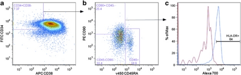
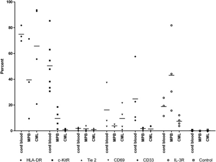
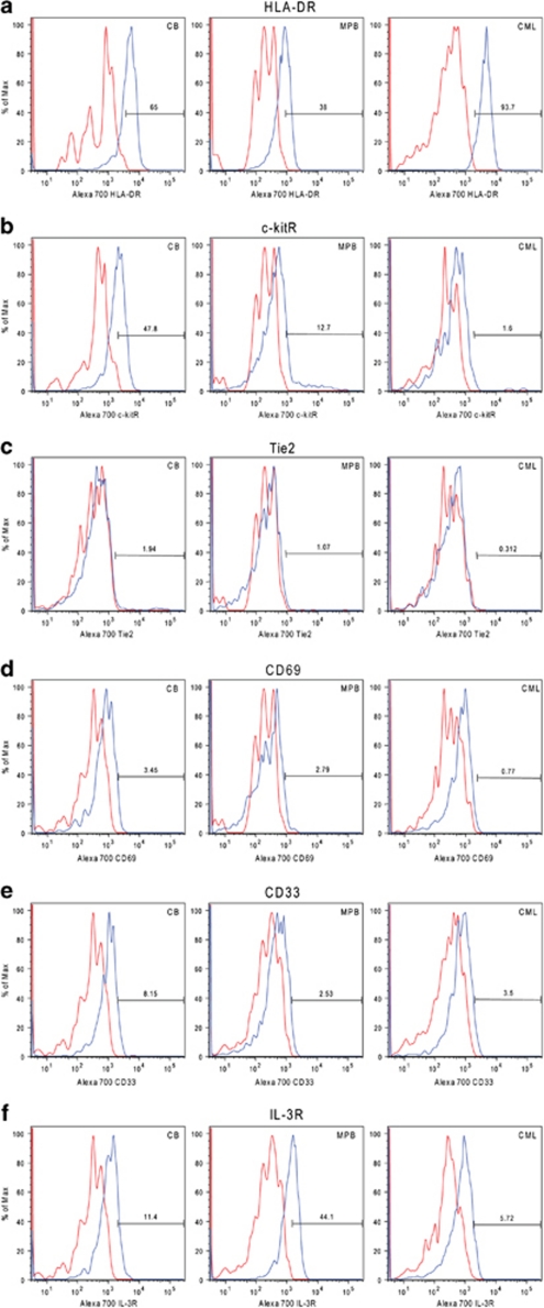
The most primitive hematopoietic stem cell (HSC)/progenitor cell (PC) population reported to date is characterized as being Lin-CD34+CD38-CD90+CD45R. We have a long-standing interest in comparing the characteristics of hematopoietic progenitor cell populations enriched from normal subjects and patients with chronic myelogenous leukemia (CML). In order to investigate further purification of HSCs and for potential targetable differences between the very primitive normal and CML stem/PCs, we have phenotypically compared the normal and CML Lin-CD34+CD38-CD90+CD45RA- HSC/PC populations. The additional antigens analyzed were HLA-DR, the receptor tyrosine kinases c-kit and Tie2, the interleukin-3 cytokine receptor, CD33 and the activation antigen CD69, the latter of which was recently reported to be selectively elevated in cell lines expressing the Bcr-Abl tyrosine kinase. Notably, we found a strikingly low percentage of cells from the HSC/PC sub-population isolated from CML patients that were found to express the c-kit receptor (<1%) compared with the percentages of HSC/PCs expressing the c-kitR isolated from umbilical cord blood (50%) and mobilized peripheral blood (10%). Surprisingly, Tie2 receptor expression within the HSC/PC subset was extremely low from both normal and CML samples. Using in vivo transplantation studies, we provide evidence that HLA-DR, c-kitR, Tie2 and IL-3R may not be suitable markers for further partitioning of HSCs from the Lin-CD34+CD38-CD90+CD45RA- sub-population.
Figures
Similar articles
-
Expression of integrin α2 receptor in human cord blood CD34+CD38-CD90+ stem cells engrafting long-term in NOD/SCID-IL2Rγ(c) null mice.Stem Cells. 2013 Feb;31(2):360-71. doi: 10.1002/stem.1282. Stem Cells. 2013. PMID: 23165626
-
Severe functional alterations in vitro in CD34(+) cell subpopulations from patients with chronic myeloid leukemia.Leuk Res. 2004 Jun;28(6):639-47. doi: 10.1016/j.leukres.2003.11.005. Leuk Res. 2004. PMID: 15120942
-
Microarray and serial analysis of gene expression analyses identify known and novel transcripts overexpressed in hematopoietic stem cells.Cancer Res. 2004 Jul 1;64(13):4434-41. doi: 10.1158/0008-5472.CAN-03-3247. Cancer Res. 2004. PMID: 15231652
-
Identification of a hierarchy of multipotent hematopoietic progenitors in human cord blood.Cell Stem Cell. 2007 Dec 13;1(6):635-45. doi: 10.1016/j.stem.2007.10.001. Cell Stem Cell. 2007. PMID: 18371405 Free PMC article.
-
Characterization and selection of benign stem cells in chronic myeloid leukemia.Haematologica. 1993 Nov-Dec;78(6):393-400. Haematologica. 1993. PMID: 8175034 Review.
Cited by
-
Preeclampsia in pregnancy affecting the stemness and differentiation potency of haematopoietic stem cell of the umbilical cord blood.BMC Pregnancy Childbirth. 2020 Jul 10;20(1):399. doi: 10.1186/s12884-020-03084-7. BMC Pregnancy Childbirth. 2020. PMID: 32650736 Free PMC article.
-
Preparation of Proper Immunogen by Cloning and Stable Expression of cDNA coding for Human Hematopoietic Stem Cell Marker CD34 in NIH-3T3 Mouse Fibroblast Cell Line.Adv Pharm Bull. 2015 Mar;5(1):69-75. doi: 10.5681/apb.2015.009. Epub 2015 Mar 5. Adv Pharm Bull. 2015. PMID: 25789221 Free PMC article.
-
Bone Marrow Mesenchymal Stem Cell-Derived Exosomal MicroRNA-126-3p Inhibits Pancreatic Cancer Development by Targeting ADAM9.Mol Ther Nucleic Acids. 2019 Jun 7;16:229-245. doi: 10.1016/j.omtn.2019.02.022. Epub 2019 Mar 1. Mol Ther Nucleic Acids. 2019. Retraction in: Mol Ther Nucleic Acids. 2022 Aug 22;29:617. doi: 10.1016/j.omtn.2022.08.024. PMID: 30925451 Free PMC article. Retracted.
-
The effects of 3D culture on the expansion and maintenance of nucleus pulposus progenitor cell multipotency.JOR Spine. 2020 Dec 8;4(1):e1131. doi: 10.1002/jsp2.1131. eCollection 2021 Mar. JOR Spine. 2020. PMID: 33778405 Free PMC article.
-
Concise Review: Chronic Myeloid Leukemia: Stem Cell Niche and Response to Pharmacologic Treatment.Stem Cells Transl Med. 2018 Mar;7(3):305-314. doi: 10.1002/sctm.17-0175. Epub 2018 Feb 8. Stem Cells Transl Med. 2018. PMID: 29418079 Free PMC article. Review.
References
-
- Smith C. Hematopoietic stem cells and hematopoiesis. Cancer Control. 2003;10:9–16. - PubMed
-
- Wang JC, Doedens M, Dick JE. Primitive human hematopoietic cells are enriched in cord blood compared with adult bone marrow or mobilized peripheral blood as measured by the quantitative in vivo SCID-repopulating cell assay. Blood. 1997;89:3919–3924. - PubMed
-
- Gothot A, Pyatt R, McMahel J, Rice S, Srour EF. Functional heterogeneity of human CD34(+) cells isolated in subcompartments of the G0/G1 phase of the cell cycle. Blood. 1997;90:4384–4393. - PubMed
-
- Schriber JR, Dejbakhsh-Jones S, Kusnierz-Glaz CR, Ginzton N, Still B, Negrin RS, et al. Enrichment of bone marrow and blood progenitor (CD34+) cells by density gradients with sufficient yields for transplantation. Exp Hematol. 1995;23:1024–1029. - PubMed
Grants and funding
LinkOut - more resources
Full Text Sources
Other Literature Sources
Molecular Biology Databases
Research Materials
Miscellaneous
