

The "quad-partite" synapse: microglia-synapse interactions in the developing and mature CNS
- PMID: 22829357
- PMCID: PMC4082974
- DOI: 10.1002/glia.22389
The "quad-partite" synapse: microglia-synapse interactions in the developing and mature CNS
Abstract
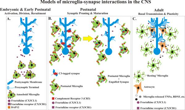
Microglia are the resident immune cells and phagocytes of our central nervous system (CNS). While most work has focused on the rapid and robust responses of microglia during CNS disease and injury, emerging evidence suggests that these mysterious cells have important roles at CNS synapses in the healthy, intact CNS. Groundbreaking live imaging studies in the anesthetized, adult mouse demonstrated that microglia processes dynamically survey their environment and interact with other brain cells including neurons and astrocytes. More recent imaging studies have revealed that microglia dynamically interact with synapses where they appear to serve as "synaptic sensors," responding to changes in neural activity and neurotransmitter release. In the following review, we discuss the most recent work demonstrating that microglia play active roles at developing and mature synapses. We first discuss the important imaging studies that have led us to better understand the physical relationship between microglia and synapses in the healthy brain. Following this discussion, we review known molecular mechanisms and functional consequences of microglia-synapse interactions in the developing and mature CNS. Our current knowledge sheds new light on the critical functions of these mysterious cells in synapse development and function in the healthy CNS, but has also incited several new and interesting questions that remain to be explored. We discuss these open questions, and how the most recent findings in the healthy CNS may be related to pathologies associated with abnormal and/or loss of neural circuits.
Copyright © 2012 Wiley Periodicals, Inc.
Figures
References
-
- Alexander A, Barres B, Stevens B. The complement system: an unexpected role in synaptic pruning during development and disease. Annual Review of Neuroscience. 2012;35:369–389. - PubMed
-
- Awasaki T, Tatsumi R, Takahashi K, Arai K, Nakanishi Y, Ueda R, Ito K. Essential role of the apoptotic cell engulfment genes draper and ced-6 in programmed axon pruning during Drosophila metamorphosis. Neuron. 2006;50:855–67. - PubMed
Publication types
MeSH terms
Grants and funding
LinkOut - more resources
Full Text Sources
