. 2013 Jan;32(1):8-17.
doi: 10.1109/TMI.2012.2209183. Epub 2012 Jul 17.

Application of micro-computed tomography with iodine staining to cardiac imaging, segmentation, and computational model development

Affiliations

Application of micro-computed tomography with iodine staining to cardiac imaging, segmentation, and computational model development

Oleg V Aslanidi et al. IEEE Trans Med Imaging. 2013 Jan.

Abstract

Micro-computed tomography (micro-CT) has been widely used to generate high-resolution 3-D tissue images from small animals nondestructively, especially for mineralized skeletal tissues. However, its application to the analysis of soft cardiovascular tissues has been limited by poor inter-tissue contrast. Recent ex vivo studies have shown that contrast between muscular and connective tissue in micro-CT images can be enhanced by staining with iodine. In the present study, we apply this novel technique for imaging of cardiovascular structures in canine hearts. We optimize the method to obtain high-resolution X-ray micro-CT images of the canine atria and its distinctive regions-including the Bachmann's bundle, atrioventricular node, pulmonary arteries and veins-with clear inter-tissue contrast. The imaging results are used to reconstruct and segment the detailed 3-D geometry of the atria. Structure tensor analysis shows that the arrangement of atrial fibers can also be characterized using the enhanced micro-CT images, as iodine preferentially accumulates within the muscular fibers rather than in connective tissues. This novel technique can be particularly useful in nondestructive imaging of 3-D cardiac architectures from large animals and humans, due to the combination of relatively high speed ( ~ 1 h/per scan of the large canine heart) and high voxel resolution (36 μm) provided. In summary, contrast micro-CT facilitates fast and nondestructive imaging and segmenting of detailed 3-D cardiovascular geometries, as well as measuring fiber orientation, which are crucial in constructing biophysically detailed computational cardiac models.


Figures

Figure 1
Figure 1
3D geometry of the canine heart reconstructed from micro-CT. A: Epicardial view. B: Endocardial view. Isosurfaces (yellow) of the X-ray intensity in micro-CT tissue images are shown, with isovalues chosen such that the epicardial and endocardial surfaces of the heart are seen clearly. Fine tissue structures, such as small blood vessels in the RV (seen in A) and the network of PMs inside the RAA (seen in B) can be resolved from the high resolution (voxel size of 36 μm) images. RAA, right atrial appendage; RV, right ventricle; PMs, pectinate muscles.
Figure 2
Figure 2
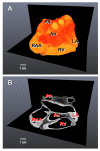
Non-segmented 3D geometry of the canine atria. A: 3D volume rendering of the dissected atria (X-ray intensity is colour-coded using a “hot metal” palette) shows the direct 3D volume renderings - this is also. Details of anatomical structures are not clearly visible, for example large aorta (Ao) is obscured by other tissues. B: Respective micro-CT images (orthogonal planes x-y and x-z) with clear inter-tissue contrast between structures, which enables segmentation of the 3D geometries (see below). Colour brightness in A and B is related to the X-ray intensity in micro-CT images: brighter colours (such as yellow in A, white in B) correspond to lower intensity/higher absorption of X-rays, which is due to higher accumulation of iodine in the respective tissues. Ao, aorta; PA, pulmonary artery; RAA, right atrial appendage; LA, left atrium, RV, right ventricle.
Figure 3
Figure 3
Segmentation of the 3D geometry of blood vessels. A: Segmented geometry of Ao (green) and PAs (red). Segmented vascular regions are reconstructed and rendered as 3D volumetric digital masks. B: Identification of Ao and PAs (dotted green and red lines) in the respective high contrast micro-CT images (orthogonal planes x-y and x-z). Similar to Fig. 2B, brighter colours in the images (e.g., white) correspond to lower intensity/higher absorption of X-rays, which is due to higher accumulation of iodine in the respective tissues. Ao, aorta; PA, pulmonary arteries.
Figure 4
Figure 4
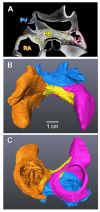
Segmentation of the 3D geometry of the canine atria. A: Identification of the RA (orange line), BB (yellow line), LA (pink line) and PVs (blue line) based on inter-tissue contrast in micro-CT images. As before, brighter colours in the latter image correspond to lower intensity/higher absorption of X-rays, which is due to higher accumulation of iodine in the respective atrial tissues. B and C: Superior and posterior views of the segmented 3D atria. Segmented atrial regions are reconstructed and rendered as 3D volumetric digital masks. 3D atrial regions are shown using same colours as lines in A. Clear structure of PMs inside the RA is seen in C. BB, Bachmann’s bundle; RA and LA, right and left atrium; PVs, pulmonary veins; PMs, pectinate muscles.
Figure 5
Figure 5
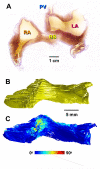
Reconstruction of fibre orientation in the BB. A: High resolution (8 μm) histological section of the sheep atria (with permission from Zhao et al., 2012). Clear arrangement of fibres along the BB can be seen. B: Segmented geometry of the BB (see also Fig. 4) used as a 3D digital mask for reconstructing fibre orientation. C: Reconstructed fibre orientation in the BB. Fibres are coloured according to their inclination angle (“rainbow” palette). Fibres are perfectly aligned in the direction along the BB, which is in agreement with histological and anatomical studies.
Figure 6
Figure 6
Reconstruction of fibre orientation in the PVs. A and B: Segmented geometry of the PV region (see also Fig. 4) used as a 3D digital mask for reconstructing fibre orientation. C and D: Reconstructed fibre orientation in the PVs. Fibres are coloured according to their inclination angle (“rainbow” palette). Fibres are mostly aligned along the PV sleeves, but their arrangement becomes more complex - with multiple changing directions - towards the LA (red arrow), which is in agreement with histological and electro-anatomical studies (Hocini et al., 2002; Zhao et al., 2012; Ho et al., 2012). A characteristic pattern of fibres (Ho et al., 2012) can be seen in the inter-pulmonary area, where circumferentially or obliquely aligned strands meet with predominantly longitudinal strands (white arrows). Posterior (A, C) and anterior (B, D) views are shown.
Figure 7
Figure 7
Reconstruction of the AVN from contrast micro-CT images. A: Identification of the AVN margins (dotted blue line) in the contrast-enhanced images. B: Segmented 3D structure of the AVN (blue). As in Figs. 3-6, the segmented region is reconstructed and rendered as a 3D volumetric digital mask. C: Anatomy of the AVN. The PNE is identified at the base of the tricuspid valve and continues anteriorly as the CN and AVB. D: Relationship of the segmented AVN to surrounding tissues seen in micro-CT images. The AVN occupies a central location in the atrioventricular septum. Ao, aorta; AS and VS, atrial and ventricular septum; PNE, posterior nodal extension; CN, compact node; AVB, atrioventricular bundle; LA and RA, left and right atrium; RV, right ventricle.

References

    1. Aslanidi OV, Colman MA, Stott J, Dobrzynski H, Boyett MR, Holden AV, Zhang H. 3D virtual human atria: a computational platform for studying clinical atrial fibrillation. Prog Biophys Mol Biol. 2011;107:156–68. - PMC - PubMed
    1. Aslanidi OV, Butters TD, Ren CX, Ryecroft G, Zhang H. Electrophysiological models for the heterogeneous canine atria: computational platform for studying rapid atrial arrhythmias. Conf Proc IEEE Eng Med Biol Soc. 2011;2011:1693–96. - PMC - PubMed
    1. Batchelor PG, Atkinson D, Hill DL, Calamante F, Connelly A. Anisotropic noise propagation in diffusion tensor MRI sampling schemes. Magn Reson Med. 2003;49:1143–51. - PubMed
    1. Benson AP, Aslanidi OV, Zhang H, Holden AV. The canine virtual ventricular wall: a platform for dissecting pharmacological effects on propagation and arrhythmogenesis. Prog Biophys Mol Biol. 2008;96:187–208. - PubMed
    1. Bordas R, Gillow K, Lou Q, Efimov IR, Gavaghan D, Kohl P, Grau V, Rodriguez B. Rabbit-specific ventricular model of cardiac electrophysiological function including specialized conduction system. Prog Biophys Mol Biol. 2011;107:90–100. - PMC - PubMed

Publication types