. 2012 Jul 24;3(4):e00193-12.
doi: 10.1128/mBio.00193-12. Print 2012.

Enterococcus faecalis produces abundant extracellular structures containing DNA in the absence of cell lysis during early biofilm formation

Affiliations

Enterococcus faecalis produces abundant extracellular structures containing DNA in the absence of cell lysis during early biofilm formation

Aaron M T Barnes et al. mBio. .

Abstract

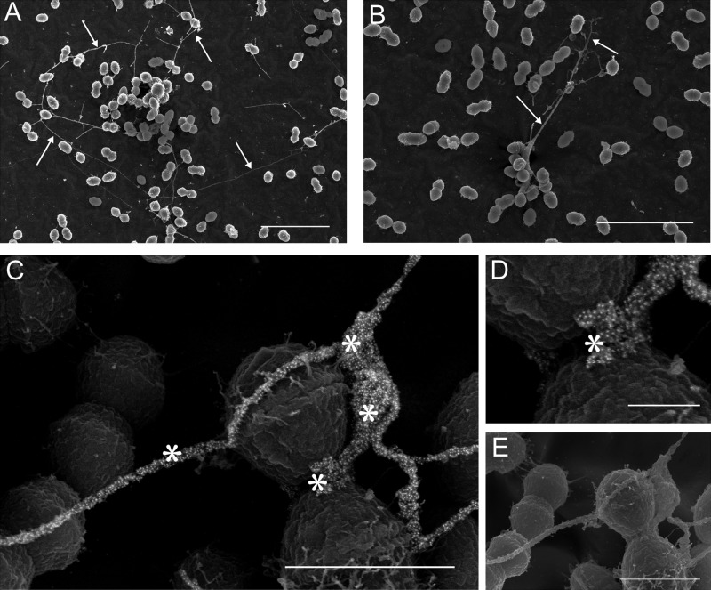
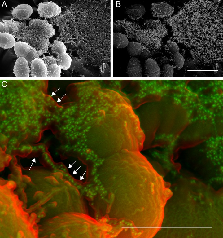
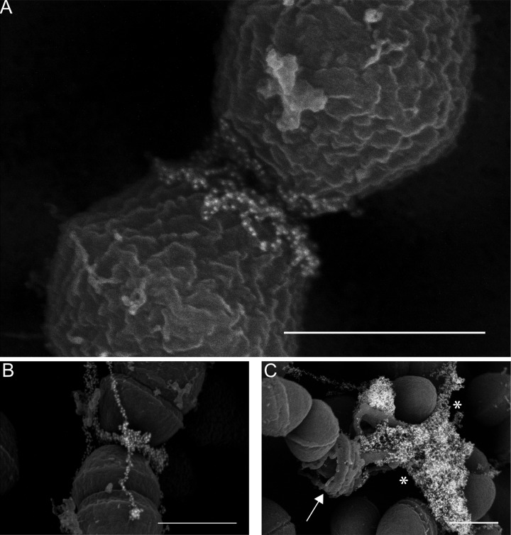
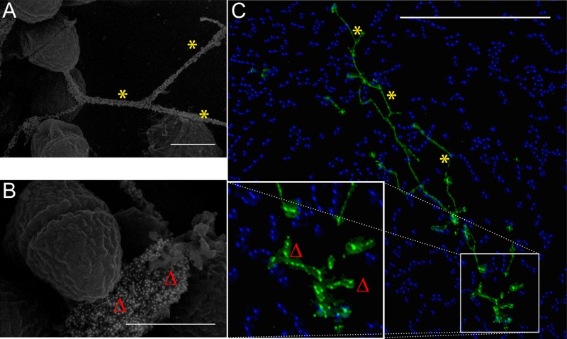
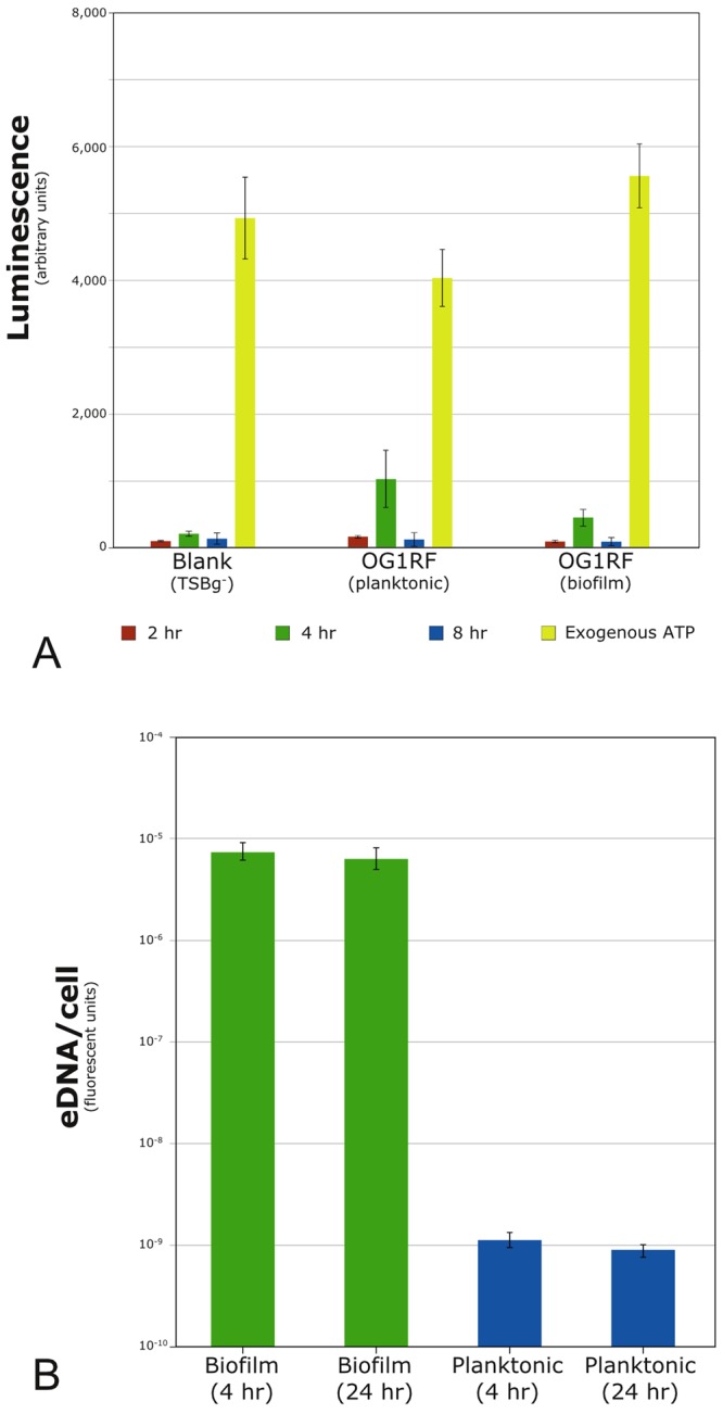
Enterococcus faecalis is a common Gram-positive commensal bacterium of the metazoan gastrointestinal tract capable of biofilm formation and an opportunistic pathogen of increasing clinical concern. Dogma has held that biofilms are slow-growing structures, often taking days to form mature microcolonies. Here we report that extracellular DNA (eDNA) is an integral structural component of early E. faecalis biofilms (≤4 h postinoculation). Combining cationic dye-based biofilm matrix stabilization techniques with correlative immuno-scanning electron microscopy (SEM) and fluorescent techniques, we demonstrate that--in early E. faecalis biofilms--eDNA localizes to previously undescribed intercellular filamentous structures, as well as to thick mats of extruded extracellular matrix material. Both of these results are consistent with previous reports that early biofilms are exquisitely sensitive to exogenous DNase treatment. High-resolution SEM demonstrates a punctate labeling pattern in both structures, suggesting the presence of an additional, non-DNA constituent. Notably, the previously described fratricidal or lytic mechanism reported as the source of eDNA in older (≥24 h) E. faecalis biofilms does not appear to be at work under these conditions; extensive visual examination by SEM revealed a striking lack of lysed cells, and bulk biochemical assays also support an absence of significant lysis at these early time points. In addition, some cells demonstrated eDNA labeling localized at the septum, suggesting the possibility of DNA secretion from metabolically active cells. Overall, these data are consistent with a model in which a subpopulation of viable E. faecalis cells secrete or extrude DNA into the extracellular matrix.

Importance: This paper reports the production of extracellular DNA during early biofilm formation in Enterococcus faecalis. The work is significant because the mechanism of eDNA (extracellular DNA) production is independent of cell lysis and the DNA is confined to well-defined structures, suggesting a novel form of DNA secretion by viable cells. Previous models of biofilm formation in enterococci and related species propose cell lysis as the mechanism of DNA release.


Figures

FIG 1
FIG 1
(A and B) Long intercellular strands (yarn structures) visible in early (4-h) E. faecalis biofilms (arrows) Bar, 5 µm. (C) Strong punctate labeling of the strands visualized by using an anti-dsDNA monoclonal antibody conjugated to an immunogold particle (asterisks). The image shows the significant eDNA component (the central area is magnified in panel D). Bar, 250 nm. (E) Surface morphology can be appreciated in the corresponding secondary electron image. Bar, 1 µm.
FIG 2
FIG 2
Thick enveloping mats of extracellular matrix (sweater structure) make up a large fraction of the E. faecalis biofilm (A, secondary electron image) and contain significant eDNA, as shown via immunogold labeling of the anti-dsDNA monoclonal antibody (B, backscatter image of the same field). Bars, 1 µm. (C) False-color SEM image showing the relationship between the surface of the ECM (red; secondary electron detector) and the immunogold-labeled anti-dsDNA antibody (green; backscatter detector). eDNA appears to be encased within another ECM component (arrows). Bar, 500 nm.
FIG 3
FIG 3
(A and B) Immuno-SEM micrographs demonstrating localization of the eDNA probe near the E. faecalis septum. (C) Endogenous lysis of cells in an older (48-h) biofilm display an entirely different morphology from that seen in early biofilms, as DNA (asterisks) is released from a ruptured cell (arrow). Bars, 500 nm.
FIG 4
FIG 4
Correlative microscopy. Though imaged using different microscopic techniques, both the SEM (A and B; bars, 500 nm) and immunofluorescent micrographs (C) show similar patterns of eDNA labeling in early E. faecalis biofilms—long intercellular strands (yarn structures [asterisks]) as well as a pericellular labeling component of the sweater matrix (Δ) (C; bar, 25 µm). Monoclonal antibody labeling of extracellular dsDNA (green, DyLight 488; blue, Hoechst 33342 highlighting individual E. faecalis cells) is shown.
FIG 5
FIG 5
Biochemical assays. (A) Luciferase-based quantification of extracellular ATP. No significant differences were found between biofilm and planktonic levels at 2, 4, or 8 h postinoculation in matched E. faecalis samples. Results are averages from four independent experiments. Only the values for the 4-h OG1RF samples (planktonic and biofilm) are above the instrument background; neither rose to statistical significance. (B) The cell-impermeant fluorescent dye Sytox green (Invitrogen) was used to label nucleic acids outside functional cells. eDNA levels were >103-fold higher under biofilm conditions than in matched planktonic controls. Exogenous lysis of a small fraction of cells (~5% by microscopy) produced a luciferase signal that saturated the instrument detector under the displayed conditions (not shown). Error bars indicate 1 SD.
FIG 6
FIG 6
Elevated membrane potentials occur in a subpopulation of biofilm cells and are correlated with significant eDNA appearance. DiOC2 crosses the cell membrane, nonspecifically labeling all cells green (A). Cells with a nonzero membrane potential (ΔΨ) preferentially accumulate the dye, which, at these higher concentrations, leads to dye molecule aggregation and the appearance of a second, red fluorescent signal (B); metabolically active cells display both green and red fluorescence. The sample was also labeled with the anti-dsDNA antibody, here conjugated to a blue fluorophore (C). Overlaying the red and blue channels (D and E) reveals that >90% of cells immediately adjacent to areas with eDNA present (asterisks) not only demonstrate viability (ΔΨ > 0) but also display membrane potentials ~25% greater than the median. Bars, 25 µm.

Similar articles

Cited by

References

    1. Flemming HC, Wingender J. 2010. The biofilm matrix. Nat. Rev. Microbiol. 8:623–633 - PubMed
    1. Lewis K. 2008. Multidrug tolerance of biofilms and persister cells. Curr. Top. Microbiol. Immunol. 322:107–131 - PubMed
    1. Whitchurch CB, Tolker-Nielsen T, Ragas PC, Mattick JS. 2002. Extracellular DNA required for bacterial biofilm formation. Science 295:1487 - PubMed
    1. Thomas VC, et al. 2009. A fratricidal mechanism is responsible for eDNA release and contributes to biofilm development of Enterococcus faecalis. Mol. Microbiol. 72:1022–1036 - PMC - PubMed
    1. Vilain S, Pretorius JM, Theron J, Brözel VS. 2009. DNA as an adhesin: Bacillus cereus requires extracellular DNA to form biofilms. Appl. Environ. Microbiol. 75:2861–2868 - PMC - PubMed

Publication types

MeSH terms

LinkOut - more resources