. 2012;8(7):e1002772.
doi: 10.1371/journal.pgen.1002772. Epub 2012 Jul 19.

Loss of ATRX, genome instability, and an altered DNA damage response are hallmarks of the alternative lengthening of telomeres pathway

Affiliations

Loss of ATRX, genome instability, and an altered DNA damage response are hallmarks of the alternative lengthening of telomeres pathway

Courtney A Lovejoy et al. PLoS Genet. 2012.

Abstract

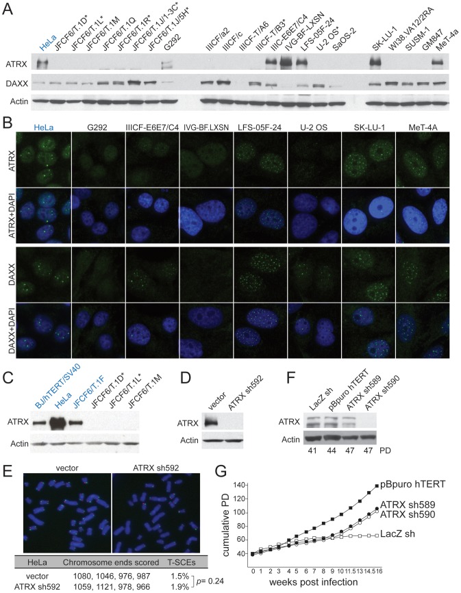
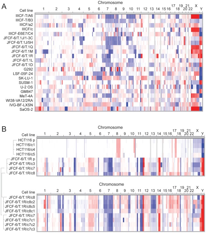
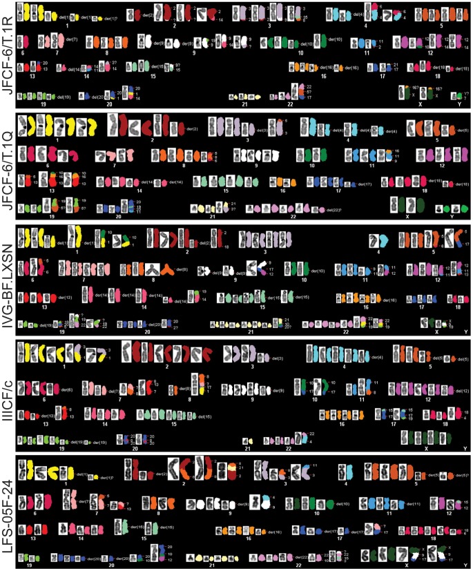
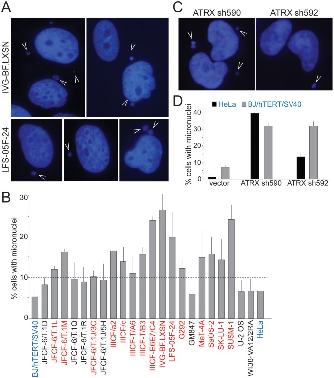
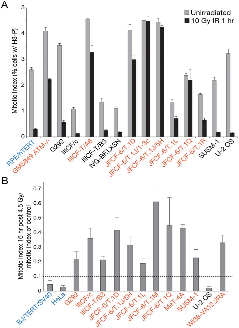
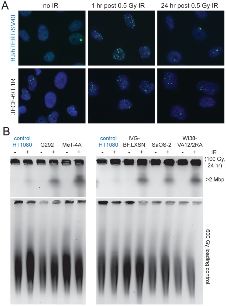
The Alternative Lengthening of Telomeres (ALT) pathway is a telomerase-independent pathway for telomere maintenance that is active in a significant subset of human cancers and in vitro immortalized cell lines. ALT is thought to involve templated extension of telomeres through homologous recombination, but the genetic or epigenetic changes that unleash ALT are not known. Recently, mutations in the ATRX/DAXX chromatin remodeling complex and histone H3.3 were found to correlate with features of ALT in pancreatic neuroendocrine cancers, pediatric glioblastomas, and other tumors of the central nervous system, suggesting that these mutations might contribute to the activation of the ALT pathway in these cancers. We have taken a comprehensive approach to deciphering ALT by applying genomic, molecular biological, and cell biological approaches to a panel of 22 ALT cell lines, including cell lines derived in vitro. Here we show that loss of ATRX protein and mutations in the ATRX gene are hallmarks of ALT-immortalized cell lines. In addition, ALT is associated with extensive genome rearrangements, marked micronucleation, defects in the G2/M checkpoint, and altered double-strand break (DSB) repair. These attributes will facilitate the diagnosis and treatment of ALT positive human cancers.


Conflict of interest statement

The authors have declared that no competing interests exist.

Figures

Figure 1
Figure 1. Deficiency in ATRX/DAXX correlates with ALT.
A, Immunoblot for ATRX and DAXX in the indicated ALT lines. ATRX was detected with A301-045 (Bethyl Labs). DAXX was detected with A301-353A (Bethyl Labs). Asterisk: cell lines with deletions in the ATRX gene (see Table 1 and Table S2). HeLa cells (blue) were used as a positive control. B, IF for ATRX and DAXX in the indicated cell lines. HeLa cells (blue) are used as a positive control. C, Immunoblot for ATRX in additional non-ALT, positive control cells (blue) relative to the indicated ALT lines. D, Immunoblot showing >90% reduction of ATRX protein in HeLa cells expressing ATRX shRNA592. E, T-SCEs, a measure for telomeric recombination, assessed by Chromosome Orientation (CO)-FISH on metaphases harvested from HeLa cells expressing vector or ATRX shRNA592. The average percentage of T-SCEs from four independent experiments is shown, with a p value derived from a two-tailed, unpaired t test. F, Immunoblot showing >90% reduction of ATRX protein in cells expressing ATRX shRNA590 but not with shRNA589. G, Growth curves showing immortalization of SV40-transformed BJ fibroblasts infected with an hTERT expressing retrovirus but no immortalization after infection with the effective (sh590) and ineffective (sh589) shRNAs to ATRX.
Figure 2
Figure 2. Copy number analysis showing extensive genome rearrangements in ALT lines.
SNP array copy number results are shown for (A) the ALT cell lines and (B) the 1st and 2nd generation subclones derived from JFCF-6/T.1R (indicated by the brackets) and subclones derived from the telomerase-positive HCT116 control. Segmented copy number data is shown for each chromosome, by genomic position in columns and by cell line in rows. The color scale ranges from red (amplification; log2 copy number ratio of 1.5) through white (neutral; 2 copies in diploid lines, log2 ratio of 0) to blue (deletion; log2 ratio of −1.5).
Figure 3
Figure 3. Abnormal karyotypes in ALT lines.
Representative metaphases from 5 cell lines showing subtetraploid karyotype with high number of rearranged chromosomes (up to 60%). Structural rearrangements are labeled as following: del(Z)- deletion of chromosome Z; der(Z) - multiple aberrations within single chromosome Z; chromosome, denoted with two or more numbers indicate rearrangement involving two or more partners.
Figure 4
Figure 4. Frequent micronucleation in ALT cells may be attributable to loss of ATRX.
A, Examples of micronuclei (arrowheads) in two ALT cell lines. Blue: DAPI stain for DNA. B, Graph showing micronucleation frequencies in the ALT cell lines and two telomerase positive controls (blue). Values are means ±SD from three experiments (>100 nuclei each). Red: cell lines with >10% micronucleation frequency (dashed line). HeLa cells were analyzed once. C, Examples of micronuclei (arrowheads) in HeLa cells expressing two independent ATRX shRNAs (see Figure 1 for immunoblots). D, Graph showing micronucleation frequencies in two telomerase positive cell lines infected with vector or the indicated ATRX shRNAs. Values are means ±SEM from two experiments (200 nuclei each).
Figure 5
Figure 5. Many ALT lines have a defect in the G2/M checkpoint initiation and/or maintenance.
A, Bar graph depicting the results of an assay for the initiation of the G2/M checkpoint at 1 hr after irradiation of the indicated cell lines. RPE/hTERT is a positive control (in blue). Mean ± SD for triplicate experiments are shown. B, Bar graph depicting the results of an assay for the maintenance of the G2/M checkpoint at 16 hr after IR. BJ/hTERT/SV40 is a positive control (in blue). Mean ± SD for triplicate experiments are shown. See Table 1 for summary. Cell lines in red have a defect in the G2/M checkpoint.
Figure 6
Figure 6. Deficiency in DSB repair in ALT lines.
A, Example of assay for 53BP1 foci in the positive control (BJ/hTERT/SV40, blue) (top) and an ALT line (bottom). Cells were treated as indicated above the panels and stained for DNA with DAPI (blue) in conjunction with IF for 53BP1 (green). Note the higher level of 53BP1 foci in non-irradiated ALT cells and incomplete DSB repair at 24 hr after IR. B, Example of PFGE-assay for DSB repair in the indicated cell lines. Cells were treated with 100 Gy IR 24 hr before harvesting and residual DSBs were evaluated based on the DNA fragments released from agarose embedded cells into a PFGE gel. Plugs were treated with 600 Gy to fragment the DNA and run in parallel to serve as a control for loading. DNA was stained with ethidium bromide after electrophoresis. HT1080 (fibrosarcoma cell line) was used as a positive control (in blue).

References

    1. Bryan TM, Englezou A, Dalla-Pozza L, Dunham MA, Reddel RR. Evidence for an alternative mechanism for maintaining telomere length in human tumors and tumor-derived cell lines. Nat Med. 1997;3:1271–1274. - PubMed
    1. Dunham MA, Neumann AA, Fasching CL, Reddel RR. Telomere maintenance by recombination in human cells. Nat Genet. 2000;26:447–450. - PubMed
    1. Bechter OE, Shay JW, Wright WE. The Frequency of Homologous Recombination in Human ALT Cells. Cell Cycle. 2004;3:457–549. - PubMed
    1. Londono-Vallejo JA, Der-Sarkissian H, Cazes L, Bacchetti S, Reddel RR. Alternative lengthening of telomeres is characterized by high rates of telomeric exchange. Cancer Res. 2004;64:2324–2327. - PubMed
    1. Cesare AJ, Griffith JD. Telomeric DNA in ALT cells is characterized by free telomeric circles and heterogeneous t-loops. Mol Cell Biol. 2004;24:9948–9957. - PMC - PubMed

Publication types

MeSH terms