. 2012;8(7):e1002816.
doi: 10.1371/journal.pgen.1002816. Epub 2012 Jul 19.

The Aspergillus nidulans MAPK module AnSte11-Ste50-Ste7-Fus3 controls development and secondary metabolism

Affiliations

The Aspergillus nidulans MAPK module AnSte11-Ste50-Ste7-Fus3 controls development and secondary metabolism

Özgür Bayram et al. PLoS Genet. 2012.

Abstract

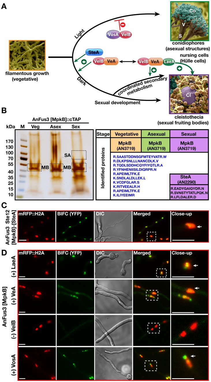
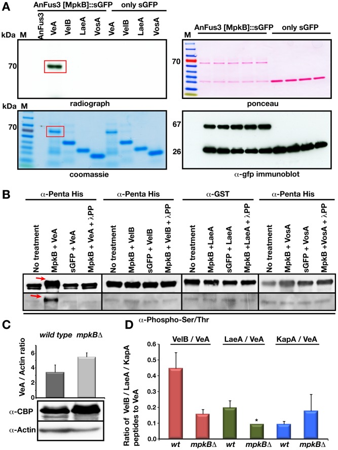
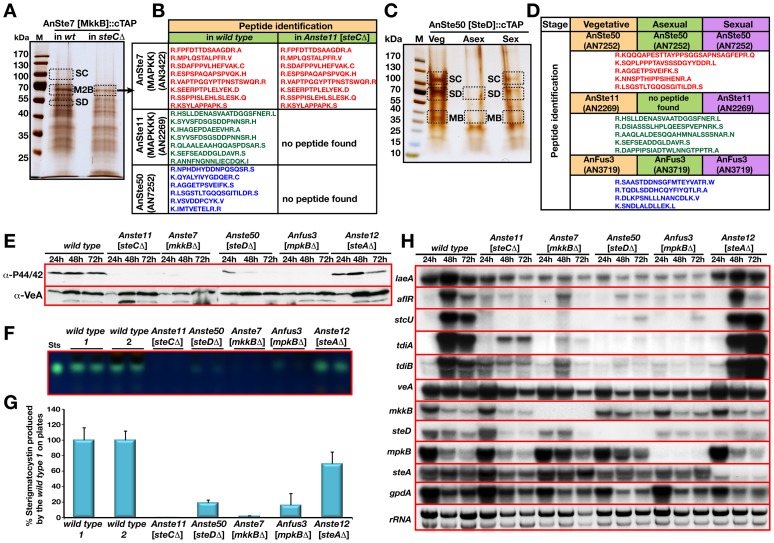
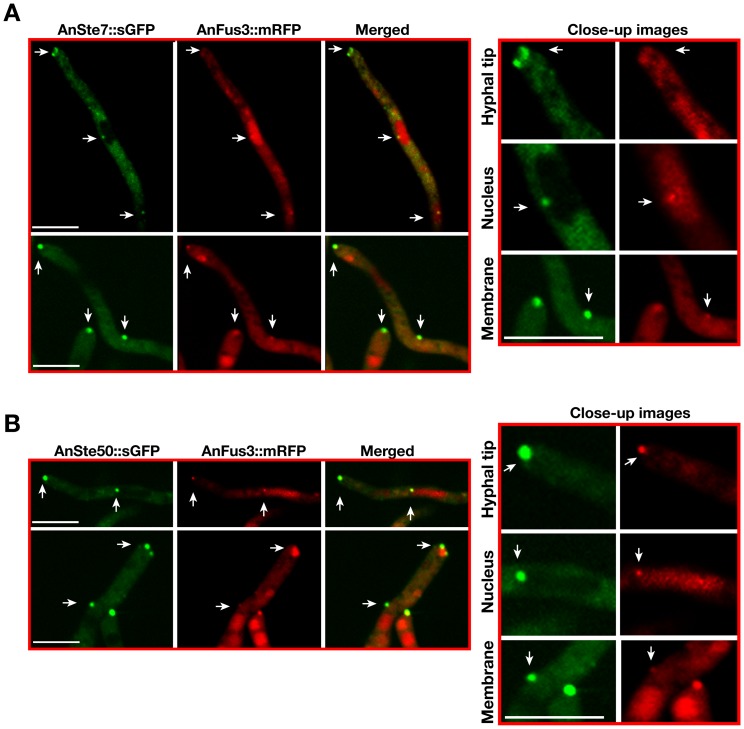
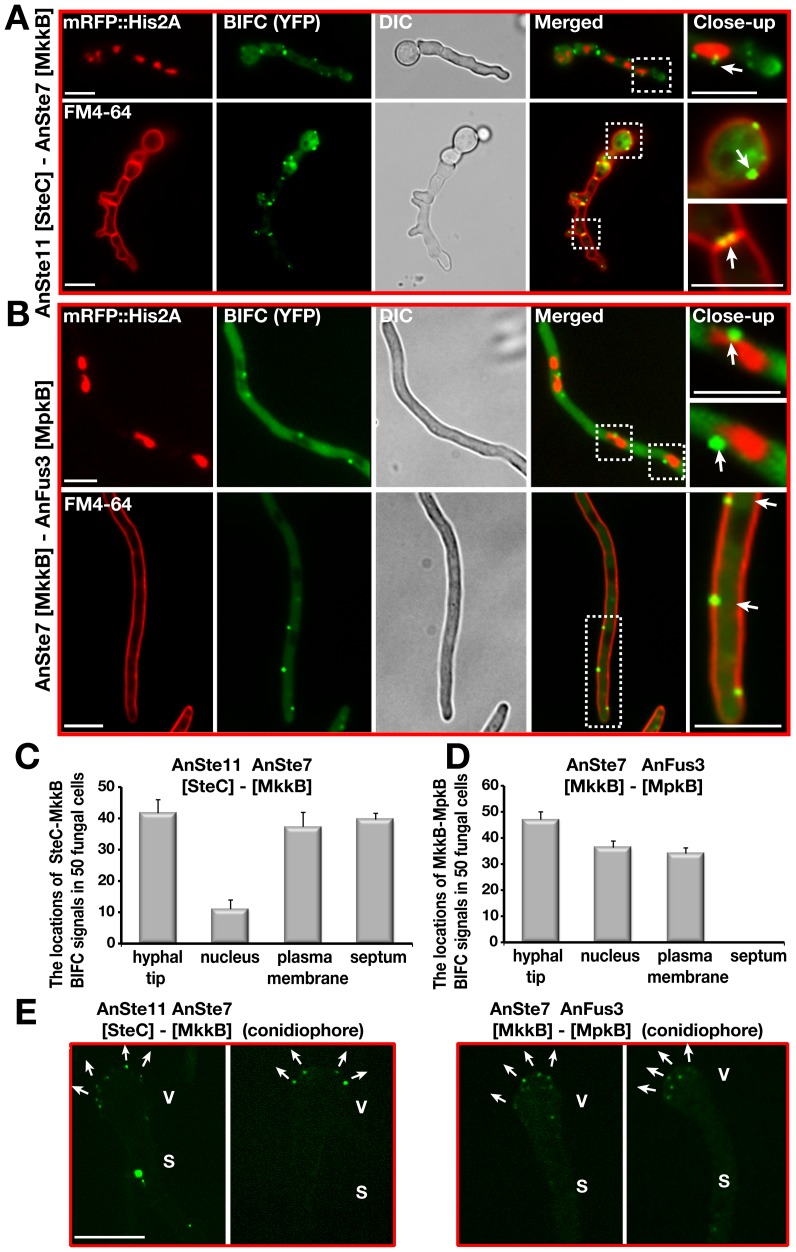
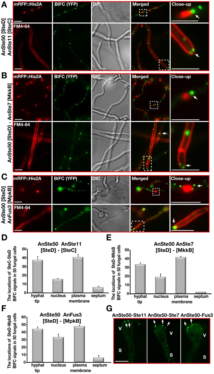
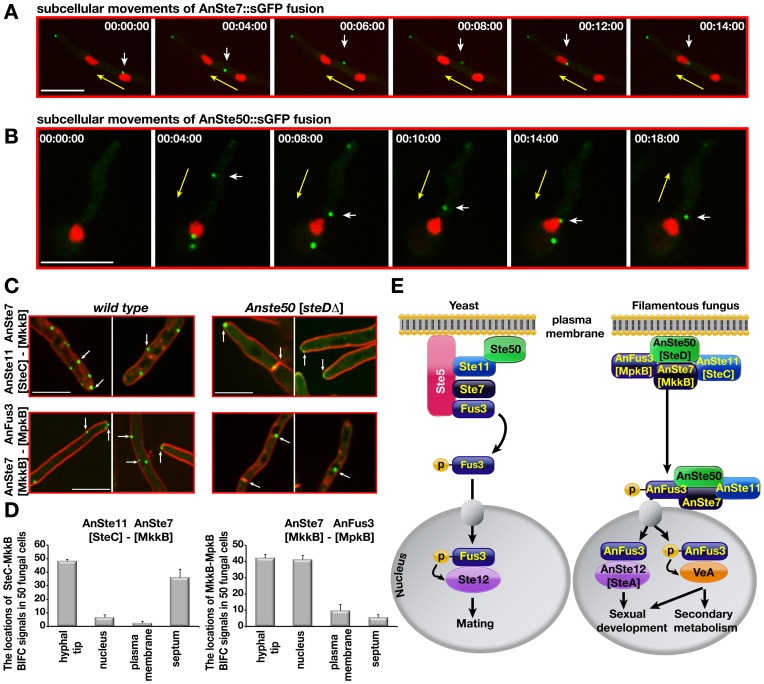
The sexual Fus3 MAP kinase module of yeast is highly conserved in eukaryotes and transmits external signals from the plasma membrane to the nucleus. We show here that the module of the filamentous fungus Aspergillus nidulans (An) consists of the AnFus3 MAP kinase, the upstream kinases AnSte7 and AnSte11, and the AnSte50 adaptor. The fungal MAPK module controls the coordination of fungal development and secondary metabolite production. It lacks the membrane docking yeast Ste5 scaffold homolog; but, similar to yeast, the entire MAPK module's proteins interact with each other at the plasma membrane. AnFus3 is the only subunit with the potential to enter the nucleus from the nuclear envelope. AnFus3 interacts with the conserved nuclear transcription factor AnSte12 to initiate sexual development and phosphorylates VeA, which is a major regulatory protein required for sexual development and coordinated secondary metabolite production. Our data suggest that not only Fus3, but even the entire MAPK module complex of four physically interacting proteins, can migrate from plasma membrane to nuclear envelope.


Conflict of interest statement

The authors have declared that no competing interests exist.

Figures

Figure 1
Figure 1. Identification of AnFus3 [MpkB] associated proteins and AnFus3 interactions with the velvet complex components.
(A) Life cycle of Aspergillus nidulans and developmental functions of AnSte12 [SteA], LaeA and velvet domain proteins. Germination of spores leads to tube-like vegetative filaments (hyphae) which become competent for environmental signals after at least 12 hours of growth. Exposure of developmentally competent hyphae to light (or aeration) leads to asexual development (conidiophores and asexual spores [conidia]) in 24 hours. VosA-VelB inhibits asexual differentiation. Incubation in dark (96 hours) induces the sexual cycle with sexual fruiting bodies (cleistothecia) which are nursed by globose Hülle cells. LaeA is required for Hülle cell formation. VelB-VeA supports sexual development together with AnSte12 [SteA]. The VelB-VeA-LaeA trimeric complex coordinates development with secondary metabolism. Co; conidia, S; stalk, Cl; cleistothecium, Hc; Hülle cells. (B) A silver stain treated 5–14% gradient SDS polyacrylamide gel of AnFus3 [MpkB]::cTAP from vegetatively, asexually (on plates, under light) and sexually (on plates, in the dark) grown cultures at 30°C for 20 hours. Identified proteins from the excised lanes (Table S1). SA; SteA-AnSte12, MB; MpkB-AnFus3. (C) AnFus3-AnSte12 interaction in vivo. N-EYFP::AnFus3 [MpkB] fusion interacts with C-EYFP fusion of AnSte12 [SteA] in the nuclei (arrow) which were visualized by a monomeric red fluorescent protein histone 2A fusion (mRFP::Histone2A). (D) Interaction partners of AnFus3 [MpkB] in BIFC. (+) indicates AnFus3 interactions with VosA and LaeA at very early stages after germination (10–12 hours) and with VeA after 24 hours of hyphal growth. (−) indicates that VelB does not interact with AnFus3. Scale bars are 10 µm.
Figure 2
Figure 2. Phosphorylation of VeA by the MAP kinase AnFus3 [MpkB] and influence of AnFus3 on the interactions of the velvet complex.
(A) In vitro phosphorylation of VeA by AnFus3 in a radioactive kinase assay. Left panels; autoradiograph of dried SDS gel run for phosphorylation reactions (30 µl of total 45 µl reaction volume), Coomassie stain of the proteins from phosphorylation reaction (10 µl of total 45 µl reaction). VeA protein in AnFus3 reaction tube is shown with red rectangle. Right panels; ponceau staining of the immunoprecipitated (immobilized to the GFP trap sepharose) AnFus3::sGFP and only sGFP protein. Immunodetection of the fusion protein and free sGFP by the α-gfp. (B) Confirmation of specific VeA phosphorylation by a non-radioactive method. All recombinant proteins (10 µg each) were treated with both AnFus3 and GFP. AnFus3 treated samples were additionaly incubated with the lambda protein phosphatase (λ-PP). Proteins were immunodetected by α-Penta His, α-GST. After AnFus3 treatment, VeA showed a 3–5 kDA molecular weight shift (red arrow) that disappeared after L-PP treatment. Only VeA treated with MAPK was recognized by P-ser/thr specific antibody. (C) Protein levels of VeA in the wild type and mpkB mutant background. VeA::cTAP signals were normalized to the internal actin levels. VeA protein levels did not change in the absence of MpkB. (D) Reduced velvet complex formation in the mpkB mutant. The VeA-associated proteins from the cultures of the wild type and mpkBΔ strains grown in the darkness sexually at 30°C for 20 hours. Three independent experiments were performed and the associated proteins were identified. The ratio of the peptides from VelB and LaeA to the VeA protein drastically reduced in the MAPK mutant, whereas alpha importin KapA interaction slightly increased. Black bars represent the standard error. *LaeA was only found in one of the three purifications in mpkBΔ strain, thus no error bar is assigned.
Figure 3
Figure 3. Loss of sexual fruiting bodies and heterokaryon formation in mkkBΔ strain lacking AnSte7.
(A) Sexual developments of a wild type, mkkBΔ, mkkB complementation (comp+), steCΔ [lacking AnSte11] strains, which were point inoculated (1×10⁴) and grown on minimal medium in dark conditions (5 days at 37°C). Small squares are the close-up stereomicroscopic images of the strains. Red arrows indicate the mature black fruiting bodies of the wild type and complementation strains and yellow arrows denote the premature nests produced by mkkB Δ and steC Δ strains. (B) Raster electron microscopy (REM) image of the strains from (A). The wild type fruiting body (cleistothecium: Cl) is surrounded by the globose Hülle cells. steC and mkkB mutants produce only dispersed groups of Hülle cells (yellow arrows) instead of mature fruiting bodies. (C) Monitoring hyphal fusions and heterokaryons via fluorescence microscopy. Strains bearing either cytoplasmic synthetic green fluorescent protein (sGFP) or nuclear red fluorescent protein fused histone 2A (mRFP) were used in different combinations. Only two wild types form green and red fluorescent combinations through hyphal fusions. (D) Isolated protoplasts from two wild types (yellow and green), steCΔ (green), mkkBΔ (yellow or green) were used for protoplast fusions as shown in combinations and plated on selective medium. wt/wt, wt/steCΔ, wt/mkkBΔ combinations produce fruiting bodies after 7–8 days, whereas mkkB Δ/mkkB Δ combination only produces nests.
Figure 4
Figure 4. Identification of AnSte7 [MkkB], and AnSte50 [SteD] associated proteins and role of MAPK pathway on secondary metabolism.
(A) A silver stained 5–14% gradient SDS polyacrylamide gel of AnSte7 [MkkB]::cTAP from the wild type and steCΔ [lacking AnSte11] background grown for vegetatively at 30°C for 20 hours. (B) Identified peptides of the proteins from the excised lanes of the wild type and steCΔ strains (Table S4 and S5). AnSte11 [SteC] (AN2269) and AnSte50 [SteD] (AN7252) were found as interactors of AnSte7 [MkkB]. (C) Interaction partners of the AnSte50::cTAP fusion from vegetatively, asexually and sexually grown cultures at 30°C for 20 hours (Table S6) (M2B; MkkB, MB; MpkB, SD; SteD, SC; SteC). (D) Identified polypeptides from the bands. (E) Monitoring of the phosphorylation status of the MpkB by phospho-p44/42 MAPK (Thr182/Tyr184) antibody in the wild type as well as pheromone pathway mutants grown for 24, 48 and 72 hours vegetatively. VeA protein levels served as loading control. 80 µg protein extract was loaded on each lane. (F) Production of secondary metabolite sterigmatocystin (ST) in the mutants of pheromone pathway, Anste11 [steCΔ], Anste50 [steDΔ], Anste7 [mkkBΔ], Anfus3 [mpkBΔ], Anste12 [steAΔ], respectively. Developed TLC plates show sterigmatocystin production. Sts; Sterigmatocystin standard. (G) Quantification of the ST production from the TLC plates. Wild type ST levels served as 100% standard. (H) Expression of the developmental, secondary metabolite genes in the pheromone pathway mutants. laeA, aflR, stcU for ST production, tdiA and tdiB for terrequinone (TQ) production and veA, mkkB, steD, mpkB, steA for developmental purposes were monitored. Strains were grown in the liquid medium for 24, 48 and 72 hours and total RNA was isolated and blotted (20 µg). Glycolytic gene gpdA expression and ethidium bromide stained rRNA was used as loading control.
Figure 5
Figure 5. Subcellular localizations of AnSte7 [MkkB], AnSte50 [SteD], and AnFus3 [MpkB]::sGFP fusions during the development of the fungus.
(A) Subcellular location of the AnSte7 [MkkB]::sGFP fusion protein in vegetatively and asexually growing hyphae. mRFP::Histone2A fusion indicates the position of the nuclei and FM4-64 stains the plasma membrane and the septa of the hyphae. Arrows indicate the accumulation of the fusion protein at hyphal tip, membrane and septa of the vegetative hyphae and metulae (M) of the conidiophores. (V) indicates the swollen vesicle part of the conidiophore. (B) Localization of AnSte50 [SteD]::sGFP fusion in the hyphal cells. SteD protein is cytoplasmic and after competence time (16 hours) it is enriched at the hyphal tip, membrane and nuclear envelope (indicated by arrows). (C) Nucleo-cytoplasmic and hyphal tip distribution of endogenously expressed AnFus3 [MpkB]::sGFP kinase fusion protein in the fungal hyphae. AnFus3 is found in the cytoplasm and nucleus, in late hours of vegetative growth (after 16 hours) accumulates at the plasma membrane and hyphal tips. (D) Presence of SteD protein at the base of the metulae and in the septa between the metulae (M) and phialides (P). Nuclear and partial septal localization of the AnFus3 protein in the asexual structures (arrows). Scale bars represent 10 µm.
Figure 6
Figure 6. Colocalizations of AnSte7 or AnSte50::sGFP with AnFus3::mRFP.
(A) Colocalizations of AnSte7 and AnFus3 proteins within the same fungal cell. White arrows indicate AnSte7::sGFP and AnFus3::mRFP colocalizations at the hyphal tip, membrane and on the nuclear envelope. (B) Colocalizations of AnSte50 and AnFus3 proteins within the same cell at the hyphal tip, plasma membrane and perinuclear district. Scale bars are 10 µm.
Figure 7
Figure 7. Confirmation of the subcellular interactions of the kinase complexes AnSte11-Ste7 and AnSte7-Fus3 by BIFC system.
(A) Interaction of N-EYFP::AnSte11 [SteC] and C-EYFP::AnSte7 [MkkB] proteins in the hyphal cells. AnSte11-Ste7 kinase complexes are located at the plasma membrane, septal connections, the hyphal tip and partially nuclear envelope. The upper panel shows the localization of the YFP signal in comparison to nuclear mRFP::Histone2A fluorescence. Lower panel displays the YFP signal emitting cells stained with membrane dye FM4-64. (B) Physical interaction of N-EYFP::AnSte7 [MkkB] with C-EYFP::AnFus3 [MpkB] proteins in the fungal cells. (C) Quantification of the subcellular locations of the AnSte11-Ste7 complexes that are often present at the hyphal tip, plasma membrane, septum and nuclear envelope. N:50 fungal cells were counted in triplicate. Standard deviations are presented as vertical bars. (D) Subcellular locations of the AnSte7-Fus3 interactions. AnSte7-Fus3 complexes hardly localize to the septum and are found more on the nuclear envelope. (E) Assembly of the AnSte11-AnSte7 and AnSte7-AnFus3 complexes on the surface of vesicles of asexual conidiophores. Arrows indicate the growth directions of the metulae initials on the vesicles. V; vesicle, S; stalk. Size of the scale bars is 10 µm.
Figure 8
Figure 8. Subcellular locations of the AnSte50-Ste11, AnSte50-Ste7, and AnSte50-Fus3 complexes in vivo.
(A) Interactions of N-EYFP::AnSte11 [SteC] and C-EYFP::AnSte50 [SteD] proteins in the fungal cells. Upper panel shows the nuclear mRFP signal and lower panel FM4-64 in comparison to the split YFP signal. (B) Interactions of AnSte50 [SteD] protein with AnSte7 [MkkB]. (C) Interactions of AnSte50 protein with AnFus3 [MpkB]. (D–F) Measurement of the subcellular locations of the AnSte50-Ste11, -Ste7, and Fus3 complexes that are frequently found in the nuclear envelope, plasma membrane, at the hypal tip and less often in the septal locations. Quantification was performed and analyzed as described in Figure 7. (G) Detection of the AnSte50-Ste11, AnSte50-Ste7, and AnSte50-Fus3 complexes on the vesicle (V) of the asexual conidiophore structures. S; stalk. Scale bars represent 10 µm.
Figure 9
Figure 9. Cellular movements of AnSte7 and AnSte50::sGFP fusions and interactions of the AnSte11-Ste7, AnSte7-Fus3 complexes in the steDΔ mutant lacking AnSte50.
(A) Cellular movements of the AnSte7::sGFP fusion. Single focal plane pictures of a time-lapse study for the AnSte7 fusion (in minutes) (Video S1). The MAPKK protein (white arrow) leaves the first nucleus; shortly after touching the membrane it reaches the second nucleus (visualized by RFP). The yellow arrow indicates movement direction. (B) AnSte50::sGFP cellular movement in both directions (Video S2). AnSte50 (white arrow) moves within the filament back to the nucleus (yellow arrow), and bounces back in the opposite direction after touching the nucleus. (C) Interactions of AnSte11-Ste7 complex occur at the hyphal tip, plasma membrane and the septa in wild type (see arrows). Membrane localization of AnSte11-Ste7 kinases drastically decreases in Anste50 [steDΔ] strain. Hyphal tip and septa localizations are unaffected. AnSte7-Fus3 interact at the hyphal tip, membrane and partially nuclear envelope. (D) Quantification of the locations of the AnSte11-Ste7 and AnSte7-Fus3 complexes in steDΔ strain. N:50 fungal cells were counted in triplicates. Standard deviations were given as vertical lines. Scale bar, 10 µm. (E) A comparative depiction of the MAPK modules and their action in the single-cell yeast and filamentous fungus. In the yeast system, (MAP3K)Ste11-(MAP2K)Ste7-(MAPK)Fus3 kinase complex assembles on the scaffold protein Ste5 which tethers the complex close to the plasma membrane. Ste50 is additionally required for membrane recruitment of the Ste11. Activation of Fus3 by Ste7 phosphorylation results in entry of the active Fus3 into the nucleus where it phosphorylates Ste12 transcription factor for mating responses. In the filamentous fungus, AnSte50 is partly responsible for the membrane attachment of the AnSte11-Ste7-Fus3 complex, which migrates to the nuclear envelope presumably to keep the AnFus3 active. Finally, AnFus3 (MpkB) is released into the nucleus where it interacts with SteA (Ste12) for hyphal fusions and sexual development. It also phosphorylates the velvet A protein, which in turn leads to activation of secondary metabolism with development.

References

    1. Marshall CJ. MAP kinase kinase kinase, MAP kinase kinase and MAP kinase. Curr Opin Genet Dev. 1994;4:82–89. - PubMed
    1. Yu JH. Regulation of development in Aspergillus nidulans and Aspergillus fumigatus. Mycobiology. 2010;38:229–237. - PMC - PubMed
    1. Roman E, Arana DM, Nombela C, Alonso-Monge R, Pla J. MAP kinase pathways as regulators of fungal virulence. Trends Microbiol. 2007;15:181–190. - PubMed
    1. Saito H. Regulation of cross-talk in yeast MAPK signaling pathways. Curr Opin Microbiol. 2010;13:677–683. - PubMed
    1. Bardwell L. A walk-through of the yeast mating pheromone response pathway. Peptides. 2004;25:1465–1476. - PubMed

Publication types

MeSH terms

Substances