. 2012;7(7):e41097.
doi: 10.1371/journal.pone.0041097. Epub 2012 Jul 19.

Cre-mediated stress affects sirtuin expression levels, peroxisome biogenesis and metabolism, antioxidant and proinflammatory signaling pathways

Affiliations

Cre-mediated stress affects sirtuin expression levels, peroxisome biogenesis and metabolism, antioxidant and proinflammatory signaling pathways

Yu Xiao et al. PLoS One. 2012.

Abstract

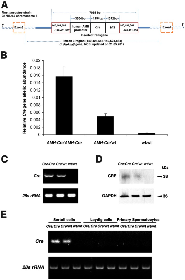
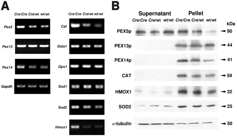
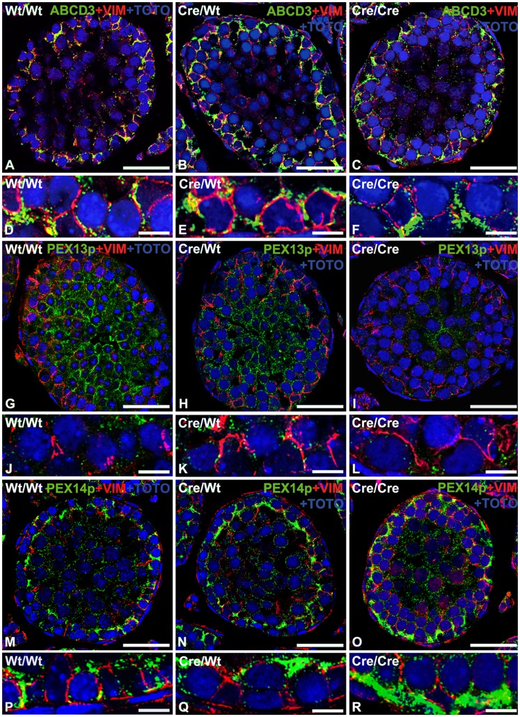
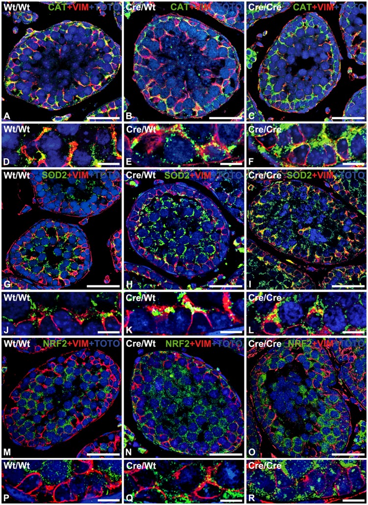
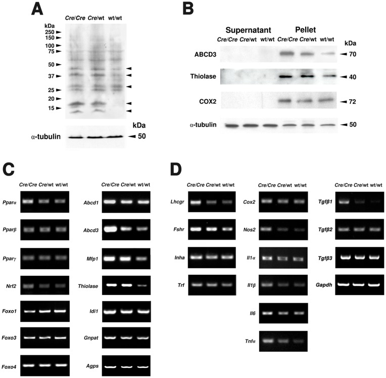
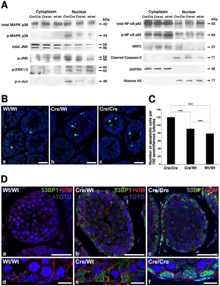
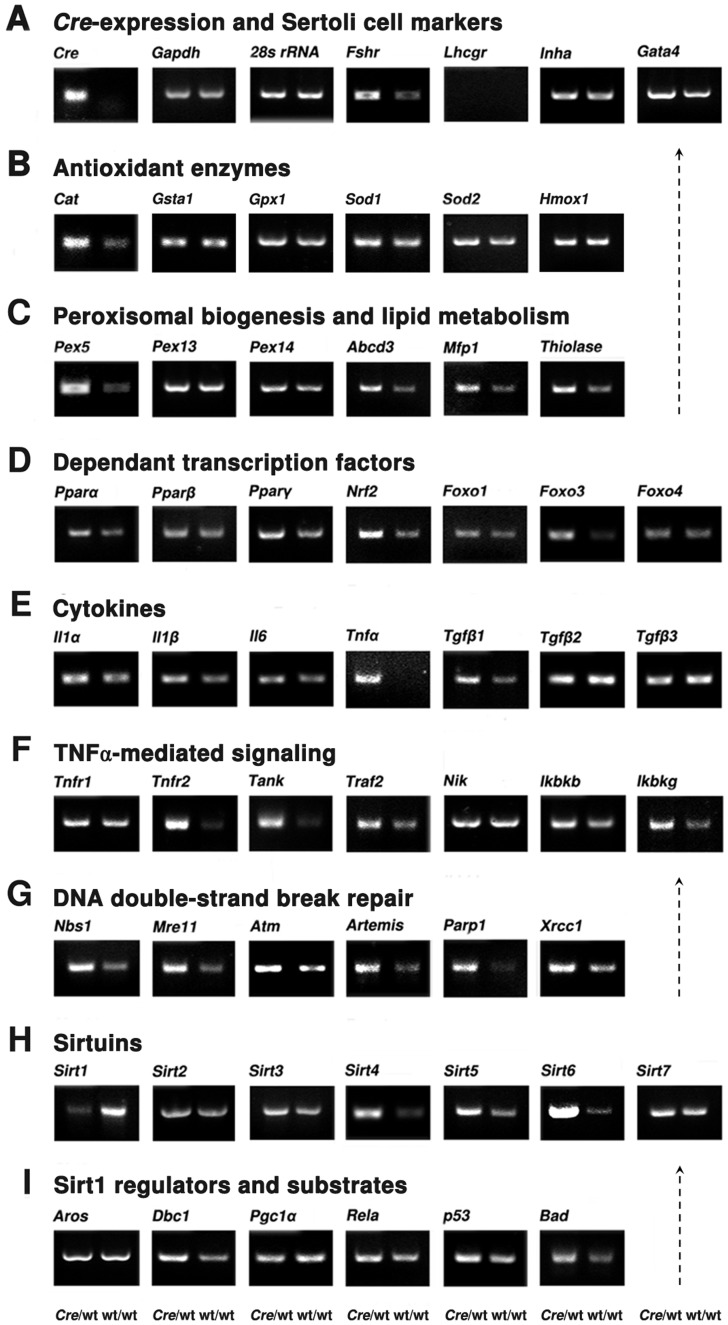
Cre-mediated excision of loxP sites is widely used in mice to manipulate gene function in a tissue-specific manner. To analyze phenotypic alterations related to Cre-expression, we have used AMH-Cre-transgenic mice as a model system. Different Cre expression levels were obtained by investigation of C57BL/6J wild type as well as heterozygous and homozygous AMH-Cre-mice. Our results indicate that Cre-expression itself in Sertoli cells already has led to oxidative stress and lipid peroxidation (4-HNE lysine adducts), inducing PPARα/γ, peroxisome proliferation and alterations of peroxisome biogenesis (PEX5, PEX13 and PEX14) as well as metabolic proteins (ABCD1, ABCD3, MFP1, thiolase B, catalase). In addition to the strong catalase increase, a NRF2- and FOXO3-mediated antioxidative response (HMOX1 of the endoplasmic reticulum and mitochondrial SOD2) and a NF-κB activation were noted. TGFβ1 and proinflammatory cytokines like IL1, IL6 and TNFα were upregulated and stress-related signaling pathways were induced. Sertoli cell mRNA-microarray analysis revealed an increase of TNFR2-signaling components. 53BP1 recruitment and expression levels for DNA repair genes as well as for p53 were elevated and the ones for related sirtuin deacetylases affected (SIRT 1, 3-7) in Sertoli cells. Under chronic Cre-mediated DNA damage conditions a strong downregulation of Sirt1 was observed, suggesting that the decrease of this important coordinator between DNA repair and metabolic signaling might induce the repression release of major transcription factors regulating metabolic and cytokine-mediated stress pathways. Indeed, caspase-3 was activated and increased germ cell apoptosis was observed, suggesting paracrine effects. In conclusion, the observed wide stress-induced effects and metabolic alterations suggest that it is essential to use the correct control animals (Cre/Wt) with matched Cre expression levels to differentiate between Cre-mediated and specific gene-knock out-mediated effects.


Conflict of interest statement

Competing Interests: The authors have declared that no competing interests exist.

Figures

Figure 1
Figure 1. Cre expression in the testis correlates with allelic abundance/genotype.
A) The consensus and integrity of inserted AMH-Cre transgene (black box) and flanking regions (red box) of Mus musculus strain C57BL/6J was confirmed by PCR-based genome walking. B) Genotyping on genomic tail DNA using quantitative real-time PCR. Values were normalized for 28s rRNA and are given as relative Cre transgene allelic abundance (average ± standard deviation). C) Semiquantitative RT-PCR analyses for the Cre expresssion and 28s rRNA on cDNAs prepared from total testicular RNA (genotypes are indicated). D) Western blot analysis of Cre and GAPDH abundance in the testis from wild type and Cre transgenic mice. 20 µg of protein were loaded per lane. E) Semiquantitative RT-PCR analyses for Cre expression and 28s rRNA on cDNAs from total RNA of microdissection samples of Sertoli cells, Leydig cells and primary spermatocytes in the testis from wild type and Cre transgenic mice.
Figure 2
Figure 2. Peroxisome biogenesis and ROS metabolism are affected by Cre recombinase expression.
A) Semiquantitative RT-PCR analyses for the genes involved in peroxisome biogenesis (Pex5, Pex13, Pex14) and ROS metabolism (Cat, Gsta1, Gpx1, Sod1, Sod2, Hmox1) on cDNAs prepared from total testicular RNA (genotypes are indicated), Gapdh as control. For abbreviation of gene names see Table 1. B) Western blot analyses of peroxisome biogenesis (PEX5p, PEX13p, PEX14p) and ROS metabolic (CAT, HMOX1, SOD2) proteins in cytosolic fractions (supernatant) and enriched organelles (pellet) of total testis samples (genotypes and protein masses are indicated). The abundance of α-tubulin was used as a loading control. 20 µg of protein were loaded per lane.
Figure 3
Figure 3. Double-immunofluorescence analysis reveals that AMH-Cre-expression induces peroxisome proliferation and membrane protein alterations.
Double immunofluorescence for peroxisomal membrane proteins (green) and the intermediate filament protein vimentin as marker for Sertoli cells (red) with counterstaining of nuclei with TOTO-3 iodide on paraffin sections from the testis of wild type (A, D, G, J, M, P), heterozygous AMH-Cre-transgenic mice (B, E, H, K, N, Q) and homozygous AMH-Cre-transgenic mice (C, F, I, L, O, R). Peroxisomes are proliferated and the lipid transporter ABCD3 is increased in individual peroxisomes in Sertoli cells in the testis of AMH-Cre-transgenic animals in comparison to wild type animals (A–F). A similar pattern is observed for PEX14p, a peroxisomal biogenesis protein with highest abundance in Sertoli cells, which was strongly induced in AMH-Cre-transgenic mice (M–R). In contrast, PEX13p, a peroxisomal biogenesis protein with highest abundance in germ cell peroxisomes is not increased in Sertoli cells and rather decreased in the germ cell population (G–L). Size of bars is: A, B, C, G, H, I, M, N, O: 25 µm; D, E, F, J, K, L, P, Q, R: 10 µm.
Figure 4
Figure 4. Immunofluorescence analysis reveals an increased antioxidative response in AMH-Cre expression Sertoli cells.
Double immunofluorescence for peroxisomal catalase (CAT) (A–F), mitochondrial superoxide dismutase 2 (SOD2) (G–L) and the redox-sensitive transcription factor NRF2 (M–R) with the intermediate filament protein vimentin as marker for Sertoli cells (red) and counterstaining of nuclei with TOTO-3 iodide on paraffin sections from the testis of wild type (A, D, G, J, M, P), heterozygous AMH-Cre-transgenic mice (B, E, H, K, N, Q) and homozygous AMH-Cre-transgenic mice (C, F, I, L, O, R). Whereas catalase is mainly present in Sertoli cells, SOD2 and NRF2 show highest abundance in spermatocytes of wild type animals. Note that all three proteins were induced in Sertoli cells of AMH-Cre-transgenic mice (see higher magnification with regions of Sertoli cells). Size of bars is: A, B, C, G, H, I, M, N, O: 25 µm; D, E, F, J, K, L, P, Q, R: 10 µm.
Figure 5
Figure 5. Alterations of gonadotropin receptors, paracrine regulators, proinflammatory genes, PPARs as well as lipid peroxidation.
A) Western blot analysis of 4-HNE-modified lysine adducts in proteins of enriched organelle pellets of the testis (genotypes are indicated). The arrows on the right indicated the altered 4-HNE-modified proteins among the three genotypes. The abundance of α-tubulin was used as a loading control. 25 µg of protein were loaded per lane. B) Western blot analyses of proteins involved in peroxisomal lipid metabolism (ABCD3), the PPARα-inducible peroxisomal β-oxidation pathway 1 (Thiolase B) and COX2-protein in enriched organelle (pellet) and cytosolic fractions (supernatant) of the testis (genotypes and protein masses are indicated). The abundance of α-tubulin was used as a loading control. 20 µg of protein were loaded per lane. C) Semiquantitative RT-PCR analyses for PPARs (Pparα-, Pparβ-, Pparγ-mRNA), redox-dependent transcription factors (Nrf2-, Foxo1-, 3-, 4-mRNA) and genes involved in peroxisomal lipid metabolism (Abcd1-, Abcd3-mRNA), PPARα-induced peroxisomal β-oxidation pathway 1 (Mfp1, Thiolase B), peroxisomal cholesterol (Idi1) and ether lipid metabolism (Gnpat, Agps). The abundance of the Gapdh mRNA was used as a loading control. D) Semiquantitative RT-PCR analyses for expression of mRNAs for gonadotropin receptors (Lhcgr, Fshr), inhibin α (Inha) and genes involved in paracrine signaling (Il1α, I1lβ, Il6, Tnfα, Tgfβ1), as well as in proinflammatory pathways (Cox2, Nos2). The expression of the Gapdh mRNA was used as a loading control.
Figure 6
Figure 6. Signaling for antioxidative response and inflammation is activated and germ cell-apoptosis is increased in AMH-Cre-mice.
A) Western blot analyses for signaling proteins of the MAP kinase family (p38, JNK, ERK1/2), c-Jun, NF-κB p65, NRF2 and caspase-3 in cytosolic and nuclear fractions of the testis (genotypes and protein masses are indicated). The abundances of GAPDH and Histone H3 were used as loading controls. 20 µg of protein were loaded per lane. B) TUNEL staining in seminiferous tubules of paraffin sections of the testis from wild type, heterozygous and homozygous AMH-Cre animals. Size of bars is 10 µm. C) Statistical analysis of TUNEL stainings revealed an increase of germ cell apoptosis in parallel to Cre allelic abundance (genotypes and number of apoptotic cells per 100 seminiferous tubules are indicated), *** p<0.001. D) Double immunofluorescence for the p53 binding protein 1 (green) and the intermediate filament protein vimentin as marker for Sertoli cells (red) with counterstaining of nuclei with TOTO-3 iodide on paraffin sections from the testis of wild type (a,d), heterozygous AMH-Cre-transgenic mice (b,e) and homozygous AMH-Cre-transgenic mice (c,f). The 53BP1 protein was induced in Sertoli cells of AMH-Cre-transgenic mice (see higher magnification with regions of Sertoli cells). Size of bars is: a-c: 25 µm; d-f: 10 µm.
Figure 7
Figure 7. Differential gene expression in the testes and by microarray-analysis were observed in isolated Sertoli cells.
Semiquantitative RT-PCR analyses for the RNA isolated from Sertoli cell cultures of AMH-Cre/Wt and Wt/Wt animals. A) Cre-transgene and gonadotropin receptors (Lhcgr, Fshr), transcription factor of Sertoli cells (Gata4), the expression levels of the Gapdh and 28s rRNA mRNAs were used as control. B) Gene expression of antioxidant enzymes (Cat, Gsta1, Gpx1, Sod1, Sod2, Hmox1). C) Genes of proteins involved in peroxisomal biogenesis and lipid metabolism (Pex5, Pex13, Pex14, Abcd3, Mfp1, Thiolase). D) Analyses of Ppar mRNAs (Pparα, Pparβ, Pparγ) and the ones for redox-dependent transcription factor (Nrf2, Foxo1, 3, 4) E) Sertoli cell cytokine mRNAs (Il1α, Il1β, Il6, Tnfα, Tgfβ1-3). F) Expression levels of components involved in the TNFα-mediated signaling pathway (Tnfr1, Tnfr2, Tank, Traf2, Nik, Ikbkb, Ikbkg). G) Expression of mRNA for proteins involved in DNA double-strand break repair (Nbs1, Mre11, Atm, Artemis, Parp1, Xrcc1). H) Gene expression levels for all mammalian sirtuins (Sirt1-7). I) Analyses of mRNA expression levels for Activator (Aros) and inhibitor of Sirt1 gene (Dbc1), as well as SIRT1 interacting partners and substrates (Pgc1α, Rela, p53, Bad).

References

    1. Gu H, Zou YR, Rajewsky K. Independent control of immunoglobulin switch recombination at individual switch regions evidenced through Cre-loxP-mediated gene targeting. Cell. 1993;73:1155–1164. - PubMed
    1. Lobe CG, Nagy A. Conditional genome alteration in mice. Bioessays. 1998;20:200–208. - PubMed
    1. Sternberg N, Hamilton D. Bacteriophage P1 site-specific recombination. I. Recombination between loxP sites. J Mol Biol. 1981;150:467–486. - PubMed
    1. Lewandoski M. Conditional control of gene expression in the mouse. Nat Rev Genet. 2001;2:743–755. - PubMed
    1. Furuta Y, Behringer RR. Recent innovations in tissue-specific gene modifications in the mouse. Birth Defects Res C Embryo Today. 2005;75:43–57. - PubMed

Publication types

MeSH terms