. 2012:2012:835731.
doi: 10.1155/2012/835731. Epub 2012 Jul 8.

Use of fluorochrome-labeled inhibitors of caspases to detect neuronal apoptosis in the whole-mounted lamprey brain after spinal cord injury

Affiliations

Use of fluorochrome-labeled inhibitors of caspases to detect neuronal apoptosis in the whole-mounted lamprey brain after spinal cord injury

Antón Barreiro-Iglesias et al. Enzyme Res. 2012.

Abstract

Apoptosis is a major feature in neural development and important in traumatic diseases. The presence of active caspases is a widely accepted marker of apoptosis. We report here the development of a method to study neuronal apoptotic death in whole-mounted brain preparations using fluorochrome-labeled inhibitors of caspases (FLICA). As a model we used axotomy-induced retrograde neuronal death in the CNS of larval sea lampreys. Once inside the cell, the FLICA reagents bind covalently to active caspases causing apoptotic cells to fluoresce, whereas nonapoptotic cells remain unstained. The fluorescent probe, the poly caspase inhibitor FAM-VAD-FMK, was applied to whole-mounted brain preparations of larval sea lampreys 2 weeks after a complete spinal cord (SC) transection. Specific labeling occurred only in identifiable spinal-projecting neurons of the brainstem previously shown to undergo apoptotic neuronal death at later times after SC transection. These neurons also exhibited intense labeling 2 weeks after a complete SC transection when a specific caspase-8 inhibitor (FAM-LETD-FMK) served as the probe. In this study we show that FLICA reagents can be used to detect specific activated caspases in identified neurons of the whole-mounted lamprey brain. Our results suggest that axotomy may cause neuronal apoptosis by activation of the extrinsic apoptotic pathway.


Figures

Figure 1
Figure 1
Schematic drawing illustrating how the FLICA method allows labeling of live cells that have activated caspases. Unbound FLICA reagent is washed out during the washing process (left side), whereas it attaches covalently to active caspases (right side).
Figure 2
Figure 2
Schematic drawing of a dorsal view of the sea lamprey brain showing the location of neuronal groups and identifiable reticulospinal neurons including giant Müller cells, a pair of Mauthner neurons (Mth), and a pair of auxiliary Mauthner neurons (mth'). Three pairs of Müller cells are identified in the caudal diencephalon (M1, M2, and M4) and one pair in the mesencephalon (M3). In the rhombencephalon, two pairs of Müller cells are identified in the anterior rhombencephalic reticular nucleus of the isthmic region (I1 and I2) and four in the middle rhombencephalic reticular nucleus or bulbar region (B1–4). Recent studies have identified additional large neurons: the I3–I6 and the B5 and B6 neurons. For abbreviations, see list. (Reproduced from [21]).
Figure 3
Figure 3
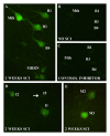
Photomicrographs of dorsal views of whole-mounted brains of larval sea lampreys show activated caspases in identified reticulospinal neurons after a complete SC transection as revealed by FAM-VAD-FMK labeling (green channel). (a) Photomicrograph of the rhombencephalon of a larval sea lamprey shows FAM-VAD-FMK labeling in the Mth, B1, B3, and B4 neurons 2 weeks after a complete SC transection. Note also the presence of labeled small-unidentified neurons of the MRRN. (b) Photomicrograph of the rhombencephalon of a larval sea lamprey shows the absence of FAM-VAD-FMK labeling in the Mth or bulbar neurons of noninjured animals. (c) Photomicrograph of the rhombencephalon of a larval sea lamprey shows the absence of FAM-VAD-FMK labeling after a complete SC transection in brains incubated with Z-VAD-FMK prior to the incubation with the FLICA reagent. (d) Photomicrograph of the rostral rhombencephalon of a larval sea lamprey shows the presence of FAM-VAD-FMK labeling in the I1 and I2 neurons 2 weeks after a complete SC transection. The arrow points to a nonlabeled I5 cell. (e) Photomicrograph of the mesencephalon/diencephalon of a larval sea lamprey shows FAM-VAD-FMK labeling in the M3 and M2 neurons 2 weeks after a complete SC transection. Rostral is up in all figures. The ventricle is at the right in all figures. For abbreviations, see list.
Figure 4
Figure 4
Photomicrographs of dorsal views of whole-mounted brains of larval sea lampreys show activated caspases in identified reticulospinal neurons after a complete SC transection as revealed by FAM-LETD-FMK labeling (green channel). (a) Photomicrograph of the rhombencephalon of a larval sea lamprey shows FAM-LETD-FMK labeling in the Mth, B1, B3, and B4 neurons 2 weeks after a complete SC transection. Note also a nonlabeled B5 cell (arrow). (b) Photomicrograph of the rostral rhombencephalon of a larval sea lamprey shows FAM-LETD-FMK labeling in the I1 and I2 neurons 2 weeks after a complete SC transection. (c) Photomicrograph of the mesencephalon/diencephalon of a larval sea lamprey shows FAM-LETD-FMK labeling in the M3 and M2 neurons 2 weeks after a complete SC transection. (d) Photomicrograph of the mesencephalon/diencephalon of a larval sea lamprey shows FAM-LETD-FMK labeling in the M1, M2, and M3 neurons 2 weeks after a complete SC transection. Rostral is up in all figures. The ventricle is at the right in all figures. For abbreviations, see list.

References

    1. Manger PR, Cort J, Ebrahim N, et al. Is 21st century neuroscience too focused on the rat/mouse model of brain function and dysfunction. Frontiers in Neuroanatomy. 2008;2, article 5 - PMC - PubMed
    1. Shifman MI, Jin L, Selzer ME. Regeneration in the lamprey spinal cord. In: Becker CG, Becker T, editors. Model Organisms in Spinal Cord Regeneration. Weinheim, Germany: Wiley-VCH Verlag GmbH; 2007. pp. 229–262.
    1. Cornide-Petronio ME, Ruiz MS, Barreiro-Iglesias A, Rodicio MC. Spontaneous regeneration of the serotonergic descending innervation in the sea lamprey after spinal cord injury. Journal of Neurotrauma. 2011;28:2535–2540. - PubMed
    1. Barreiro-Iglesias A. Targeting ependymal stem cells in vivo as a non-invasive therapy for spinal cord injury. DMM Disease Models and Mechanisms. 2010;3(11-12):667–668. - PubMed
    1. Barreiro-Iglesias A. ‘Evorego’: studying regeneration to understand evolution, the case of the serotonergic system. Brain, Behavior and Evolution. 2012;79:1–3. - PubMed