

Foetal haemoglobin, erythrocytes containing foetal haemoglobin, and hematological features in congolese patients with sickle cell anaemia
- PMID: 22830000
- PMCID: PMC3398577
- DOI: 10.1155/2012/105349
Foetal haemoglobin, erythrocytes containing foetal haemoglobin, and hematological features in congolese patients with sickle cell anaemia
Abstract
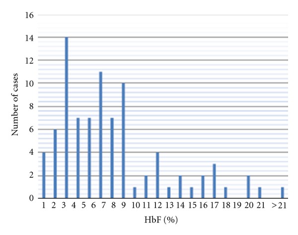
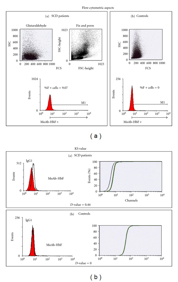
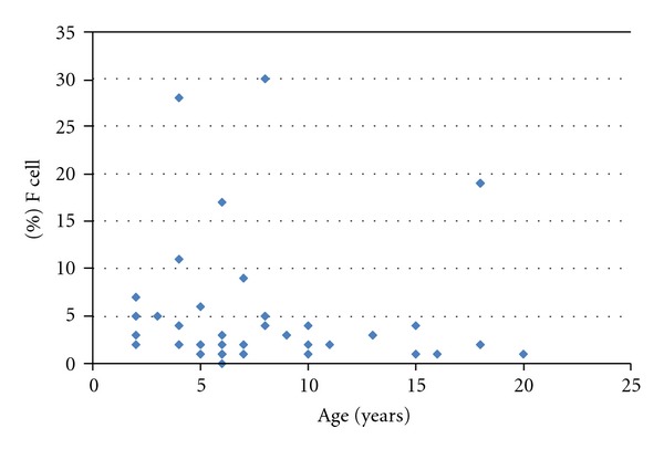
High HbF levels and F cells are correlated with reduced morbidity and mortality in sickle cell disease (SCD). This paper was designed to determine the HbF and F cells levels in Congolese sickle cell anemia (SCA) patients in order to determine their impact on the expression of SCD. Population and Method. HbF levels were measured in 89 SCA patients (mean age 11.4 yrs) using a standard HPLC method. F cell quantitation was done in a second group of SCA patients (n = 42, mean age 8.9 yrs) and compared with a control group (n = 47, mean age 5 yrs). F cells were quantified by a cytofluorometric system (MoAb-HbF-FITC; cut off at 0.5%). Results. The mean value of HbF was 7.2% ± 5.0 with heterogeneous distribution, most patients (76%) having HbF < 8%. Mean values of F-cells in SCA patients and control group were 5.4% ± 7.6 (median: 2.19%; range 0,0-30,3%) and 0.5% ± 1.6 (median 0.0, range 0-5.18), respectively. SCA patients with F cells >4.5% developed less painful crisis and had higher percentage of reticulocytes. Conclusion. Congolese SCA patients displayed low levels of HbF and F-cells that contribute to the severity of SCD.
Figures
Similar articles
-
Foetal Haemoglobin and Disease Severity in Nigerian Children with Sickle Cell Anaemia.Mediterr J Hematol Infect Dis. 2017 Nov 1;9(1):e2017063. doi: 10.4084/MJHID.2017.063. eCollection 2017. Mediterr J Hematol Infect Dis. 2017. PMID: 29181140 Free PMC article.
-
FOETAL HAEMOGLOBIN (HbF) STATUS IN ADULT SICKLE CELL ANAEMIA PATIENTS IN IBADAN, NIGERIA.Ann Ib Postgrad Med. 2010 Jun;8(1):30-3. doi: 10.4314/aipm.v8i1.63955. Ann Ib Postgrad Med. 2010. PMID: 25161472 Free PMC article.
-
Foetal haemoglobin and disease severity in sickle cell anaemia patients in Kampala, Uganda.BMC Blood Disord. 2012 Sep 7;12:11. doi: 10.1186/1471-2326-12-11. BMC Blood Disord. 2012. PMID: 22958547 Free PMC article.
-
Discovering the genetics underlying foetal haemoglobin production in adults.Br J Haematol. 2009 May;145(4):455-67. doi: 10.1111/j.1365-2141.2009.07650.x. Epub 2008 Mar 2. Br J Haematol. 2009. PMID: 19344402 Review.
-
Mechanism of action of hydroxyurea in the management of sickle cell anemia in adults.Semin Hematol. 1997 Jul;34(3 Suppl 3):15-21. Semin Hematol. 1997. PMID: 9317197 Review.
Cited by
-
Factors Associated with Growth Retardation in Children Suffering from Sickle Cell Anemia: First Report from Central Africa.Anemia. 2017;2017:7916348. doi: 10.1155/2017/7916348. Epub 2017 Jan 30. Anemia. 2017. PMID: 28250985 Free PMC article.
-
Foetal Haemoglobin and Disease Severity in Nigerian Children with Sickle Cell Anaemia.Mediterr J Hematol Infect Dis. 2017 Nov 1;9(1):e2017063. doi: 10.4084/MJHID.2017.063. eCollection 2017. Mediterr J Hematol Infect Dis. 2017. PMID: 29181140 Free PMC article.
-
Protective BCL11A and HBS1L-MYB polymorphisms in a cohort of 102 Congolese patients suffering from sickle cell anemia.J Clin Lab Anal. 2018 Jan;32(1):e22207. doi: 10.1002/jcla.22207. Epub 2017 Mar 23. J Clin Lab Anal. 2018. PMID: 28332727 Free PMC article.
-
High prevalence of individuals with low concentration of fetal hemoglobin in F-cells in sickle cell anemia in Tanzania.Am J Hematol. 2016 Aug;91(8):E323-4. doi: 10.1002/ajh.24390. Epub 2016 May 11. Am J Hematol. 2016. PMID: 27085091 Free PMC article. No abstract available.
-
Correlation between the Lactate Dehydrogenase Levels with Laboratory Variables in the Clinical Severity of Sickle Cell Anemia in Congolese Patients.PLoS One. 2015 May 6;10(5):e0123568. doi: 10.1371/journal.pone.0123568. eCollection 2015. PLoS One. 2015. PMID: 25946088 Free PMC article.
References
-
- Elion J, Labie D. Bases physiopathologiques moléculaires et cellulaires du traitement de la drépanocytose. Hématologie. 1996;2(6):499–510.
-
- Labie D, Elion J. Généthique et physiopathologie de la drépanocytose. In: Girot R, Begué P, Galactéros F, editors. La Drépanocytose. Paris, France: John Libbey Eurotext; 2003. pp. 1–11.
-
- Platt OS, Brambilla DJ, Rosse WF, et al. Mortality in sickle cell disease—life expectancy and risk factors for early death. The New England Journal of Medicine. 1994;330(23):1639–1644. - PubMed
-
- Thein SL, Menzel S. Discovering the genetics underlying foetal haemoglobin production in adults. British Journal of Haematology. 2009;145(4):455–467. - PubMed
-
- Maier-Redelsperger M, Bardakdjlan-Michau J, Neonato MG, Girot R. Diagnostic biologique des syndromes drépanocytaires. In: Girot R, Begué P, Galacteros F, editors. La Drépanocytose. Paris, France: Ed John Libbey Eurotext; 2003. pp. 13–29.
LinkOut - more resources
Full Text Sources
