

Unusual foreign bodies in the orofacial region
- PMID: 22830058
- PMCID: PMC3399346
- DOI: 10.1155/2012/191873
Unusual foreign bodies in the orofacial region
Abstract
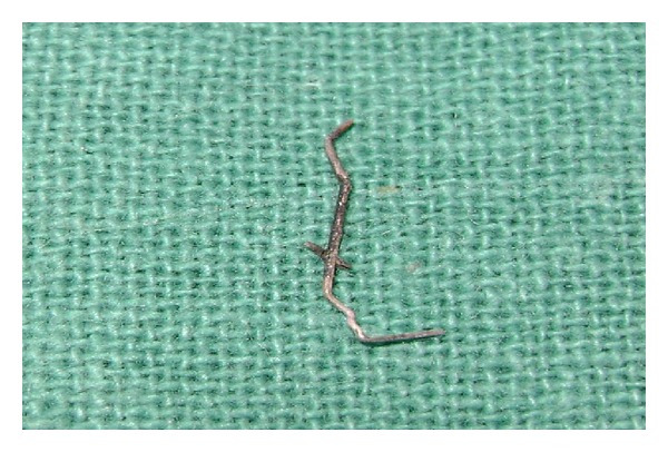
Foreign bodies may be deposited in the oral cavity either by traumatic injury or iatrogenically. Among the commonly encountered iatrogenic foreign bodies are restorative materials like amalgam, obturation materials, broken instruments, needles, and so forth. The discovery of foreign bodies in the teeth is a special situation, which is often diagnosed accidentally. Detailed case history, clinical and radiographic examinations are necessary to come to a conclusion about the nature, size, location of the foreign body, and the difficulty involved in its retrieval. It is more common to find this situation in children as it is a well-known fact that children often tend to have the habit of placing foreign objects in the mouth. Sometimes the foreign objects get stuck in the root canals of the teeth, which the children do not reveal to their parents due to fear. These foreign objects may act as a potential source of infection and may later lead to a painful condition. This paper discusses the presence of unusual foreign bodies-a tip of the metallic compass, stapler pin, copper strip, and a broken sewing needle impregnated in the gingiva and their management.
Figures


Similar articles
-
Foreign objects in teeth: retrieval and management.J Indian Soc Pedod Prev Dent. 2009 Jul-Sep;27(3):179-83. doi: 10.4103/0970-4388.57100. J Indian Soc Pedod Prev Dent. 2009. PMID: 19841551
-
Endodontic management of an unusual foreign body in a maxillary central incisor.J Conserv Dent. 2013 Sep;16(5):474-6. doi: 10.4103/0972-0707.117496. J Conserv Dent. 2013. PMID: 24082582 Free PMC article.
-
Foreign body in root canals of two adjacent deciduous molars: a case report.Int J Clin Pediatr Dent. 2013 Jan;6(1):38-9. doi: 10.5005/jp-journals-10005-1184. Epub 2013 Apr 26. Int J Clin Pediatr Dent. 2013. PMID: 25206186 Free PMC article.
-
Foreign bodies.Radiographics. 2003 May-Jun;23(3):731-57. doi: 10.1148/rg.233025137. Radiographics. 2003. PMID: 12740473 Review.
-
Management of Accidental and Iatrogenic Foreign Body Injuries to Heart- Case Series.J Clin Diagn Res. 2017 Mar;11(3):PE01-PE04. doi: 10.7860/JCDR/2017/23847.9336. Epub 2017 Mar 1. J Clin Diagn Res. 2017. PMID: 28511449 Free PMC article. Review.
Cited by
-
Embedded fingernail in the gingival sulcus of a boy.BMJ Case Rep. 2020 Jun 7;13(6):e235746. doi: 10.1136/bcr-2020-235746. BMJ Case Rep. 2020. PMID: 32513771 Free PMC article. No abstract available.
-
Clinical management and retrieval of foreign body inclusion in a primary tooth: a case report.J Med Case Rep. 2025 Aug 7;19(1):396. doi: 10.1186/s13256-025-05468-9. J Med Case Rep. 2025. PMID: 40775656 Free PMC article.
-
Impression material mass retained in the mucobuccal fold.Case Rep Dent. 2014;2014:416965. doi: 10.1155/2014/416965. Epub 2014 Jun 29. Case Rep Dent. 2014. PMID: 25061529 Free PMC article.
-
Traumatic Impaction of Unusual Foreign Body in a 10-year-old Boy's Mouth: A Case Report.Int J Clin Pediatr Dent. 2020 Jul-Aug;13(4):433-436. doi: 10.5005/jp-journals-10005-1799. Int J Clin Pediatr Dent. 2020. PMID: 33149421 Free PMC article.
-
Computer-Controlled Local Anesthesia Complication: Surgical Retrieval of a Broken Dental Needle in Noncooperative Autistic Paediatric Patient.Case Rep Dent. 2020 Nov 10;2020:6686736. doi: 10.1155/2020/6686736. eCollection 2020. Case Rep Dent. 2020. PMID: 33224533 Free PMC article.
References
-
- Ayer AA, Levin MP. Self-mutilating behaviours involving the oral cavity. Journal Blanton. 1974;29(1):4–7. - PubMed
-
- Blanton PL, Hurt WC, Largent MD. Oral factitious injuries. Journal of Periodontology. 1977;48(1):33–37. - PubMed
-
- Stewart DJ. Minor self inflicted injuries to the gingivae. Gingivitis artefacta minor. Journal of Clinical Periodontology. 1976;3(2):128–132. - PubMed
-
- Blanton PL, Hurt WC, Largent MD. Oral factitious injuries. Journal of Periodontology. 1977;48(1):33–37. - PubMed
-
- Pattison GL. Self-inflicted gingival injuries: literature review and case report. Journal of Periodontology. 1983;54(5):299–304. - PubMed
LinkOut - more resources
Full Text Sources
