

A rare root canal configuration of maxillary second molar: a case report
- PMID: 22830061
- PMCID: PMC3399371
- DOI: 10.1155/2012/767582
A rare root canal configuration of maxillary second molar: a case report
Abstract
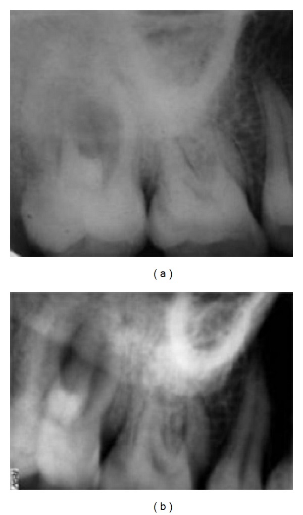
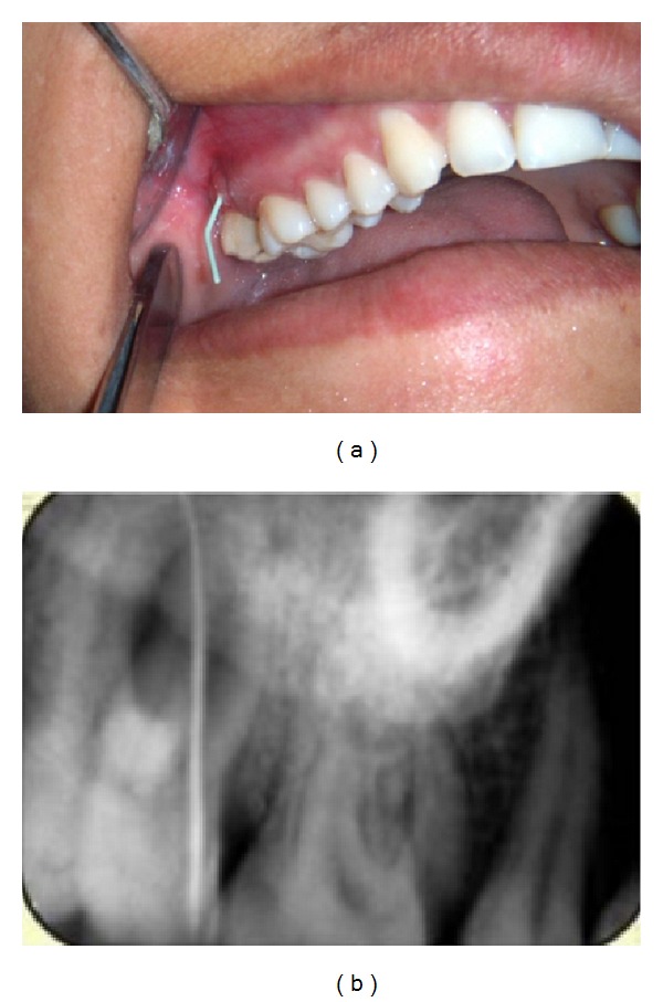
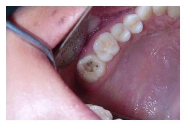
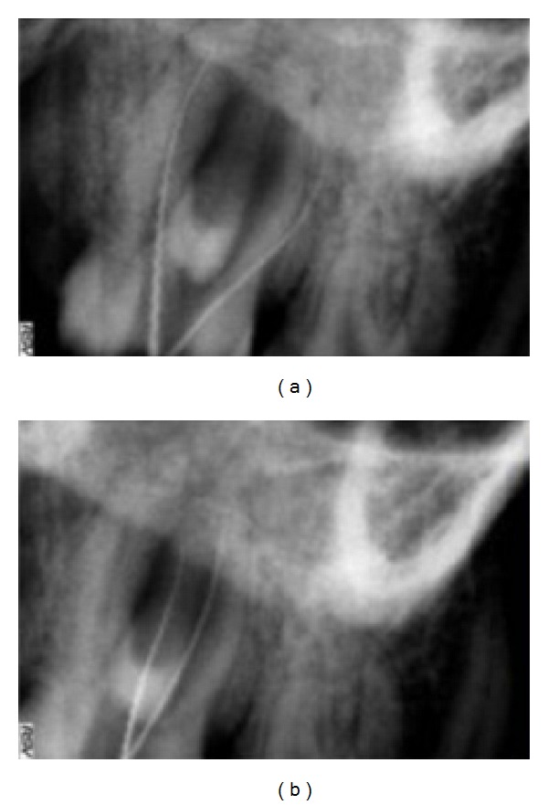
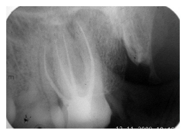
A thorough knowledge of root canal morphology is a prerequisite for the endodontic therapy. The maxillary molars, especially the second molars, have the most complicated root canal system in permanent dentition. There are many variations in canal number and configuration in maxillary molars. Treatment may be unsuccessful because the dentist may fail to recognize the unusual canal configuration. The present paper describes a case of a right maxillary second molar with a canal configuration rarely reported in the literature. The tooth had four roots with four root canals, two individual palatal roots (mesiopalatal and distopalatal) with their own separate canals. The mesiobuccal and distobuccal root had normal anatomy. This paper may intensify the complexity of maxillary molar variation and is intended to reinforce clinician's awareness of the rare morphology of root canals.
Figures
References
-
- Grossman LI, Oliet S, Rio CE. Principles of endodontic treatment. In: Grossman LI, editor. Endodontic Practice. 11th edition. Philadelphia, Pa, USA: Lea & Febiger; pp. 132–144.
-
- Slowey RR. Radiographic aids in the detection of extra root canals. Oral Surgery Oral Medicine and Oral Pathology. 1974;37(5):762–772. - PubMed
-
- Malagnino V, Gallottini L, Passariello P. Some unusual clinical cases on root anatomy of permanent maxillary molars. Journal of Endodontics. 1997;23(2):127–128. - PubMed
-
- Stone LH, Stroner WF. Maxillary molars demonstrating more than one palatal root canal. Oral Surgery Oral Medicine and Oral Pathology. 1981;51(6):649–652. - PubMed
-
- Fogel HM, Peikoff MD, Christie WH. Canal configuration in the mesiobuccal root of the maxillary first molar: a clinical study. Journal of Endodontics. 1994;20(3):135–137. - PubMed
LinkOut - more resources
Full Text Sources
