. 2012 Aug;18(4):753-60.
doi: 10.1017/S1431927612000542. Epub 2012 Jul 26.

Swept field laser confocal microscopy for enhanced spatial and temporal resolution in live-cell imaging

Affiliations

Swept field laser confocal microscopy for enhanced spatial and temporal resolution in live-cell imaging

Manuel Castellano-Muñoz et al. Microsc Microanal. 2012 Aug.

Abstract

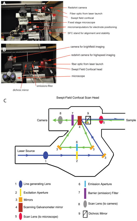
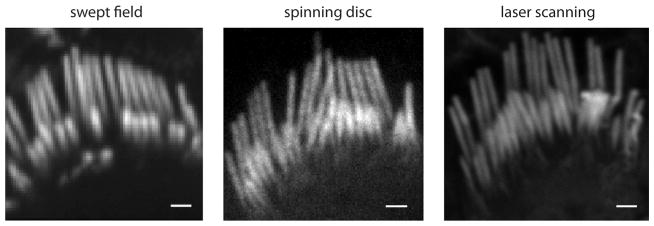
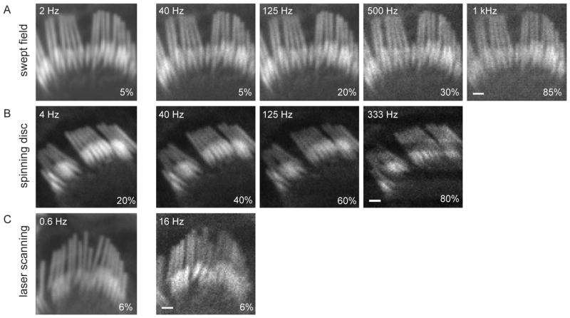
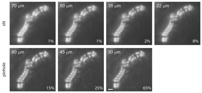
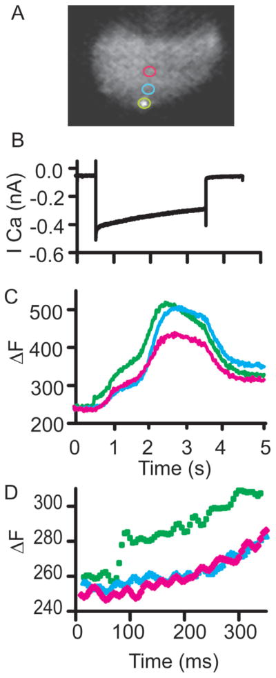
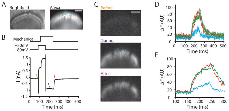
Confocal fluorescence microscopy is a broadly used imaging technique that enhances the signal-to-noise ratio by removing out of focal plane fluorescence. Confocal microscopes come with a variety of modifications depending on the particular experimental goals. Microscopes, illumination pathways, and light collection were originally focused upon obtaining the highest resolution image possible, typically on fixed tissue. More recently, live-cell confocal imaging has gained importance. Since measured signals are often rapid or transient, thus requiring higher sampling rates, specializations are included to enhance spatial and temporal resolution while maintaining tissue viability. Thus, a balance between image quality, temporal resolution, and tissue viability is needed. A subtype of confocal imaging, termed swept field confocal (SFC) microscopy, can image live cells at high rates while maintaining confocality. SFC systems can use a pinhole array to obtain high spatial resolution, similar to spinning disc systems. In addition, SFC imaging can achieve faster rates by using a slit to sweep the light across the entire image plane, thus requiring a single scan to generate an image. Coupled to a high-speed charge-coupled device camera and a laser illumination source, images can be obtained at greater than 1,000 frames per second while maintaining confocality.


Figures

Figure 1
Figure 1
SFC and electrophysiological recording station. A: Presents a photograph of the recording station with key elements highlighted. The SFC system is integrated into an electrophysiology recording station using an optical rail based support system. B: Presents a higher power view of the assembly, again highlighting the major components. A separate camera is used for the bright-field image needed for electrophysiological recordings. C: Presents the optical light path for excitation (blue) and emission (green) pathways. Again key features are labeled. Significant is the lack of moving parts that ensures stability while at the same time enhancing speed of imaging.
Figure 2
Figure 2
Comparison of SFC with spinning disc and laser scanning confocal systems. Images show stereocilia from rat inner hair cell bundles labeled with Alexa Fluor 488-phalloidin using a 100× 1.4 NA oil immersion objective. The SFC image is the average of 40 frames obtained at 40 fps using a 60 μm pinhole at 120 nm/pixel. The spinning disc image was obtained in a Yokogawa CSU10 using a 50 μm pinhole with a 2 s exposure at 46 nm/pixel. The LSCM image was obtained with a Zeiss LSM 5 Exciter for a total of 120 s at 10 nm/pixel. Scale = 1 μm.
Figure 3
Figure 3
Comparison of different sampling rates in three confocal systems. A: A hair bundle was imaged with the SFC system at different acquisition rates (2 to 1,000 fps) using a 50 μm slit. The laser intensity, depicted in the lower right corner of each panel, had to be increased for higher frequencies to obtain equivalent illumination intensities. B: Another hair bundle was imaged with the CSU10 Yokogawa spinning disc system at different sampling rates (4 to 333 fps) using a 50 μm pinhole. The maximal rate in our spinning disc microscope was 360 fps. Scan line inhomogeneity observed at higher rates is due to the inability of our spinning disc head to change its rotation speed to have integer scans of the image during our camera exposure time. C: Another hair bundle was imaged with the LSM 5 Exciter laser scanning confocal system at two acquisition rates. The image was set to 80 × 80 pixels for the sake of comparison. The fastest frequency allowed by the LSCM was 16 fps using 4 μs dwelling time. Scale = 1 μm.
Figure 4
Figure 4
Comparison of a hair bundle SFC section imaged with different slits and pinhole sizes. Rat hair cell bundle was fixed and stained with phallodin-Alexa Fluor 488. Images were acquired at 40 fps during 500 ms using different slit or pinhole sizes. Slit widths closer to the Airy limit improved resolution. Contrast was improved by using pinholes to the detriment of light exposure. The laser intensity is depicted in the lower right corner of each panel. Scale = 1 μm.
Figure 5
Figure 5
Calcium imaging of depolarization induced calcium entry using SFC imaging. A: Presents images with colored rings indicating regions sampled. The SFC image is a single frame of a hair cell with a synapse labeling peptide where a single synapse is in focus. B: Presents the calcium current. Cesium based intracellular solutions were used with apamin (100 nM) external to isolate the calcium currents. C: Provides the change in fluorescence observed at the color coded spots indicated in image A. Note the multiple components in the SFC time course. D: Provides an expanded time view to illustrate the value of temporal resolution as seen in the green trace.
Figure 6
Figure 6
Method used to localize the site of mechanotransduction channels in inner hair cell stereocilia. A: Presents the bright-field hair bundle image and the Alexa Fluor 594-filled stereocilia for identification of all stereocilia. B: Provides the stimulus above and the hair cell current responses below. The stimulus protocol first depolarizes hair bundles to reduce inward driving force for calcium while the hair bundle is moving. The hair bundle is deflected with a picospritzer to activate mechanotranducer channels, and then the cell is hyperpolarized while channels are open in order to image calcium entry. Arrows indicate time points in image C where images were obtained. C: Presents calcium images, Fluo4ff was the calcium indicator, at time points indicated in image B. The colored circles are the stereocilia where fluorescence was tracked in time (D). Scale bar represents 2 μm. D: Plots the fluorescence against time to illustrate the rapid increase in calcium levels for the second and third row but not the first. E: An expanded time view of the onset to better represent the different time courses.

References

    1. Beurg M, Fettiplace R, Nam JH, Ricci AJ. Localization of inner hair cell mechanotransducer channels using high-speed calcium imaging. Nat Neurosci. 2009;12:553–558. - PMC - PubMed
    1. Gasparini S, Losonczy A, Chen X, Johnston D, Magee JC. Associative pairing enhances action potential backpropagation in radial oblique branches of CA1 pyramidal neurons. J Physiol. 2007;580:787–800. - PMC - PubMed
    1. Gratton E, vandeVen MJ. Chapter 5: Laser sources for confocal microscopy. In: Pawley JB, editor. Handbook of Biological Confocal Microscopy. 3. New York: Springer; 2006. pp. 80–125.
    1. Inoue S. Chapter 1: Foundations of confocal scanned imaging in light microscopy. In: Pawley JB, editor. Handbook of Biological Confocal Microscopy. 3. New York: Springer; 2006. pp. 1–16.
    1. Minsky M. Memoir on inventing the confocal microscope. Scanning. 1988;10:128–138.

Publication types